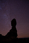
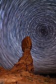
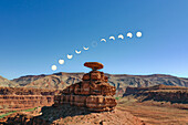
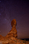
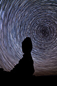
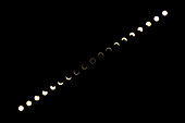
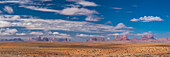
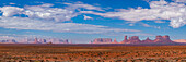
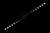
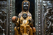
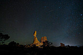
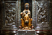
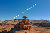
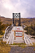
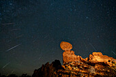
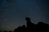
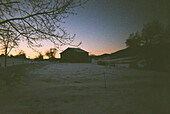
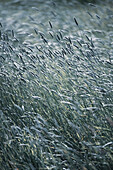
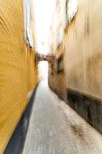
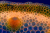
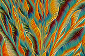
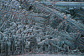
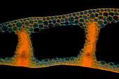
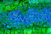
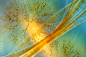
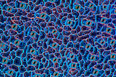
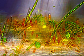
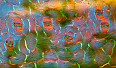
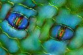
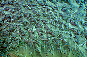
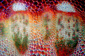
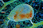
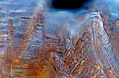
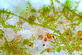
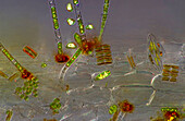
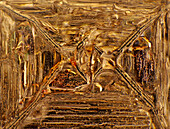
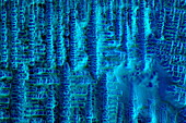
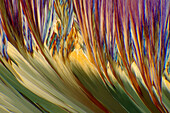
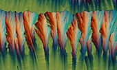
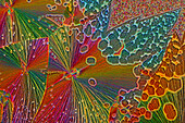
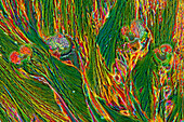
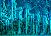
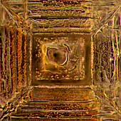
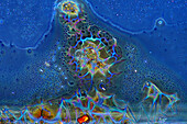
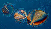
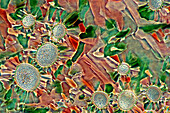
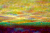
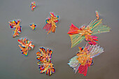
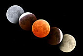
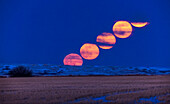
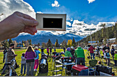
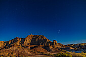
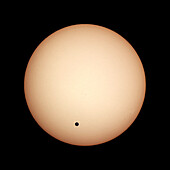
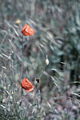
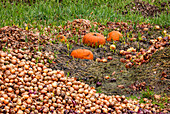
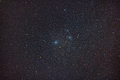
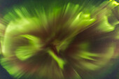
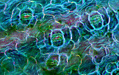
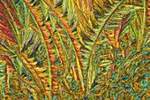
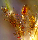
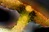
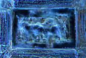
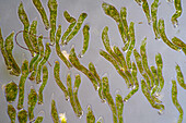
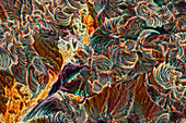
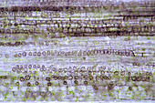
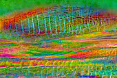
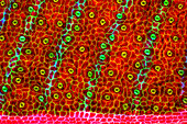
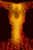
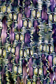
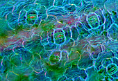
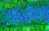
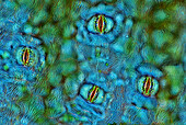
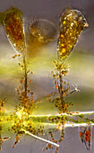
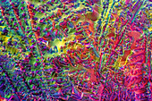
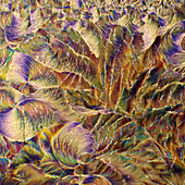
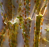
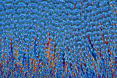
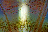
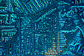
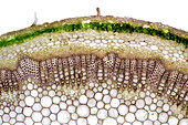
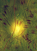
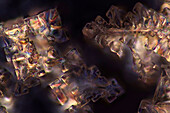
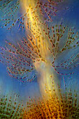
14092114 - Geminid Meteor Shower over Balanced Rock in Arches National Park in Utah. Composite image shows 5 faint meteorites over a 2-hour period.14092067 - Star trails circling the North Star over Balanced Rock in Arches National Park in winter in Utah. Composite image of 180 20-second exposures over one hour.14090103 - Composite image of Mexican Hat Rock in Utah, double-exposed with the annular eclipse on 14 October 2023. Mexican Hat Rock was very near the center of the path of peak annularity. FIltered images of the eclipse were added to the unfiltered image of Mexican Hat Rock taken prior to the eclipse.14089709 - Geminid Meteor Shower over Balanced Rock in Arches National Park in Utah. Composite image shows 5 faint meteorites over a 2-hour period.14089523 - Star trails circling the North Star over Balanced Rock in Arches National Park in winter in Utah. Composite image of 180 20-second exposures over one hour.14089304 - Geminid Meteor Shower over Balanced Rock in Arches National Park in Utah. Composite image shows 23 meteorites over a 2-hour period.14088967 - Composite image of the annular solar eclipse on 14 November 2023. Utah, USA.14126511 - A tattered American flag with the image of a Native American in traditional dress in the Monument Valley Navajo Tribal Park in Arizona.14125894 - Panoramic view of Monument Valley from the northeast. L-R: Three Sisters at the left end of Mitchell Mesa, East Mitten, Merrick Butte, West Mitten, Gray Whiskers Butte, Mitchell Butte, Sentinal Mesa, Big Indian Chief, Castle Butte, Bear & Rabbit, Stagecoach, King on the Throne, Brigham's Tomb, Eagle Mesa. The border between Arizona & Utah is between Mitchell Butte & Sentinal Mesa, almost in the very center of the image, wtih Utah at right.14123929 - Panoramic view of Monument Valley from the northeast. L-R: Three Sisters at the left end of Mitchell Mesa, East Mitten, Merrick Butte, West Mitten, Gray Whiskers Butte, Mitchell Butte, Sentinal Mesa, Big Indian Chief, Castle Butte, Bear & Rabbit, Stagecoach, King on the Throne, Brigham's Tomb, & Eagle Mesa at far right behind. The border between Arizona & Utah is between Mitchell Butte & Sentinal Mesa, almost in the very center of the image, wtih Utah at right.14091380 - Composite image of the annular solar eclipse on 14 November 2023. Utah, USA. The sun at peak annularity has been enlarged for artistic effect.14091212 - The Black Virgin is a highly regarded sculptural image of the Virgin Mary with the Child, kept in the Benedictine abbey of Santa Maria de Montserrat, Monistrol de Montserrat, Barcelona, Catalonia, Spain14089881 - Geminid Meteor Shower over Balanced Rock in Arches National Park in Utah. Composite image shows 10 meteorites over a 2-hour period.14089635 - Vaporettos Gondolas, with tourists, on the San Marco gondola stop next to the Basilica de Santa Maria della Salud, Venice, UNESCO, Veneto, Italy, Europe. Cityscape image of Grand Canal in Venice, with Santa Maria della Salute Basilica in the background.14089535 - The Black Virgin is a highly regarded sculptural image of the Virgin Mary with the Child, kept in the Benedictine abbey of Santa Maria de Montserrat, Monistrol de Montserrat, Barcelona, Catalonia, Spain14091548 - The Black Virgin is a highly regarded sculptural image of the Virgin Mary with the Child, kept in the Benedictine abbey of Santa Maria de Montserrat, Monistrol de Montserrat, Barcelona, Catalonia, Spain14090951 - Composite image of Mexican Hat Rock in Utah, double-exposed with the annular eclipse on 14 October 2023. Mexican Hat Rock was very near the center of the path of peak annularity. FIltered images of the eclipse were added to the unfiltered image of Mexican Hat Rock taken prior to the eclipse.71452521 - Vertical image of old bridge in Torres del Paine National Park, forbidden by "no pasar", highlights abandoned atmosphere and conservation of wild nature, Chile, South America 14091785 - Geminid Meteor Shower over Balanced Rock in Arches National Park in Utah. Composite image shows 23 meteorites over a 2-hour period.14089987 - Geminid Meteor Shower over Balanced Rock in Arches National Park in Utah. Composite image shows 24 meteorites over a 2-hour period.14088765 - Geminid Meteor Shower over Balanced Rock in Arches National Park in Utah. Composite image shows 24 meteorites over a 2-hour period.14130807 - Analog image of sunset landscape in Orante, Huesca, Spain71433471 - Color image of grass stems.71433469 - Multiple exposure image of a alley in Bruges, Belgium.71433393 - Fairy tale image of the rooftops and spires in medieval Bruges as seen from the side of one of the inner city canals.13925580 - The image presents Anemone sylvestris stalk in transversal cross-section, photographed through the microscope in polarized light at a magnification of 200X\n13925536 - The image presents crystallized paracetamol, photographed through the microscope in polarized light at a magnification of 100X\n13925533 - The image presents crystallized ammonium chloride photographed through the microscope in polarized light at a magnification of 100X\n13925525 - The image presents Carex sp. leaf in transversal cross-section, photographed through the microscope in polarized light at a magnification of 100X\n13925473 - The image presents stomata in Spathiphyllum leaf epidermis, photographed through the microscope in polarized light at a magnification of 100X\n13925385 - The image presents Fragilaria sp., a kind of diatoms against Batrachospermum, a kind of red algae, photographed through the microscope in polarized light at a magnification of 200X\n13925332 - The image presents stomata in hosta leaf epidermis, photographed through the microscope in polarized light at a magnification of 100X\n13925278 - The image presents various tiny algae settled on Lemna sp. root, photographed through the microscope in polarized light at a magnification of 400X. On the right are visible diatoms closed in a special protecting case.\n13925207 - The image presents stomata in Spathiphyllum leaf epidermis, photographed through the microscope in polarized light at a magnification of 400X\n13925161 - The image presents red wine photographed through the microscope in polarized light at a magnification of 100X\n13925148 - The image presents stomata in Spathiphyllum leaf epidermis, photographed through the microscope in polarized light at a magnification of 400X\n13925117 - The image presents crystallized soy sauce, photographed through the microscope in polarized light at a magnification of 100X\n13925085 - The image presents vascular bundles in senecio stalk, photographed through the microscope in polarized light at a magnification of 200X\n13925083 - The image presents Utricularia trap, a kind of carnivorous plant, photographed through the microscope in polarized light and dark field, at a magnification of 100X\n13925079 - The image presents crystallized mixture of kitchen salt and erythritol, photographed through the microscope in polarized light at a magnification of 100X\n13925044 - The image presents various tiny algae settled on Lemna sp. root, photographed through the microscope in polarized light at a magnification of 200X\n13925041 - The image presents various tiny algae settled on Lemna sp. root, photographed through the microscope in polarized light at a magnification of 200X\n13925035 - The image presents crystallized mixture of kitchen salt and erythritol, photographed through the microscope in polarized light at a magnification of 100X\n13925027 - The image presents crystallized soy sauce, photographed through the microscope in polarized light at a magnification of 100X\n13925010 - The image presents read leaf in transversal cross-section, photographed through the microscope in polarized light at a magnification of 100X\n13924998 - The image presents crystallized resorcinol, photographed through the microscope in polarized light at a magnification of 100X\n13924862 - The image presents a single crystal of recrystallized kitchen salt, photographed through the microscope in polarized light at a magnification of 200X\n13924844 - The image presents crystallized mixture of urea and paracetamol, photographed through the microscope in polarized light at a magnification of 100X\n13924827 - The image presents crystallized soy sauce, photographed through the microscope in polarized light at a magnification of 100X\n13924810 - The image presents crystallized soy sauce, photographed through the microscope in polarized light at a magnification of 100X\n13924754 - The image presents crystallized mixture of malic acid and hydroquinone photographed through the microscope in polarized light at a magnification of 100X\n13924619 - The image presents crystallized tartaric acid, photographed through the microscope in polarized light at a magnification of 100X\n13924584 - The image presents crystallized soy sauce, photographed through the microscope in polarized light at a magnification of 100X\n13924550 - The image presents a single crystal of recrystallized kitchen salt, photographed through the microscope in polarized light at a magnification of 200X\n13924465 - The image presents crystallized silver nitrate, photographed through the microscope in polarized light at a magnification of 100X\n13924437 - The image presents crystallized sulfur, photographed through the microscope in polarized light at a magnification of 100X\n13924395 - The image presents crystallized tartaric acid, photographed through the microscope in polarized light at a magnification of 100X\n13924337 - The image presents tissues in nettle stalk in longitudinal cross section, photographed through the microscope in polarized light at a magnification of 100X\n13924325 - The image presents crystallized mixture of malic acid, salicylic acid and acetanilid, photographed through the microscope in polarized light at a magnification of 100X\n13899961 - Composite image digitally created in Photoshop of images taken during October 27, 2004 total lunar eclipse, from Alberta Canada. Images taken through 5-inch apo refractor at f/6 with Canon Digital Rebel camera at ISO 200.13898786 - The rising of the Full Moon on March 9, 2020, (sometimes known as the Worm Moon) with a deer in the foreground, and in a composite of images of the Moon taken 3 minutes apart. The sky and foreground come from the first image with the Moon on the horizon. The Moon was into cloud for the last exposure.13898410 - The partial solar eclipse of October 23, 2014 as seen from Jasper, Alberta, at a public event in Centennial Park as part of the annual Dark Sky Festival. This is a single-exposure image showing the scene near mid-eclipse with telescopes from volunteers from the Royal Astronomical Society of Canada, and the mostly clear skies above with the crescent Sun visible through the handheld polymer solar filter.13898054 - The International Space Station (ISS) flying away to the east over the moonlit badlands formations at Dinosaur Provincial Park, Alberta, on July 12, 2022, just before local midnight. The image frames the stars of Cassiopeia (upper left), Perseus (at left), Andromeda (centre) and Pegasus (at right). A couple of other fainter satellites are also in the image. Light from the almost Full Moon illuminates the sky blue and foreground a warm colour.13897726 - Transit of Venus, June 8, 2004, from Luxor, Egypt. A single image taken near mid-transit. Taken with a Sony DSC-V1 digital camera shooting afocally through a 40mm eyepiece and on a 90mm apochromatic refractor, equatorially mounted and driven. Shot thru a Baader solar filter, which gives a white Sun. Yellow coloration added in Photoshop.71433476 - Desaturated image of poppy flowers.71425495 - Focus stacking image of onions dumped in large numbers in a field and some Hokkaido, affluent society in Germany13998751 - The star clusters Messier 46 (left) and Messier 47 (right) in Puppis,in a wide-field telephoto image simulating the feld of view of binoculars. The clusters NGC 2423 (above M47) and Mel71 (top) are also in the frame. The little planetary NGC 2438 embedded in M46 just shows up as a green dot.13998668 - A fish-eye lens image of auroral curtains and rays converging toward the zenith overhead at centre,with the camera looking straight up from the parking lot of the Churchill Northern Studies Centre. Taken February 19,2023. The Big Dipper is at centre.13925579 - The image presents stomata in Spathiphyllum leaf epidermis, photographed through the microscope in polarized light at a magnification of 200X\n13925520 - The image presents crystallized callus remover, photographed through the microscope in polarized light at a magnification of 100X.\n13925423 - The image presentstwo suctorians ( a kind of ciliate) and tiny diatoms, photographed through the microscope in polarized light at a magnification of 200X\n13925392 - The image presents reed stalk in transversal cross-section, photographed through the microscope in polarized light at a magnification of 200X\n13925377 - The image presents a single crystal of recrystallized salt, photographed through the microscope in polarized light at a magnification of 100X\n13925372 - The image presents a single crystal of recrystallized kitchen salt, photographed through the microscope in polarized light at a magnification of 200X\n13925367 - The image presents Ophrydium sp. ( a kind of colonial ciliates), photographed through the microscope in polarized light at a magnification of 100X\n13925342 - The image presents crystallized malic acid, photographed through the microscope in polarized light at a magnification of 100X\n13925338 - The image presents crystallized soy sauce, photographed through the microscope in polarized light at a magnification of 100X\n13925287 - The image presents tissues in nettle stalk in longitudinal cross-section, photographed through the microscope in polarized light at a magnification of 100X\n13925265 - The image presents nettle tissues in the stalk in longitudinal cross-section, photographed through the microscope in polarized light at a magnification of 100X\n13925174 - The image presents crystallized tartaric acid, photographed through the microscope in polarized light at a magnification of 100X\n13925104 - The image presents diptera eggs, photographed through the microscope in polarized light at a magnification of 100X\n13925100 - The image presents stomata in Stromanthe sp. leaf epidermis, photographed through the microscope in polarized light at a magnification of 100X\n13925093 - The image presents a single vascular bundle in Carex sp. stalk, photographed through the microscope in polarized light and dark field at a magnification of 200X\n13925078 - The image presents stomata in lily leaf epidermis, photographed through the microscope in polarized light at a magnification of 200X\n13925070 - The image presents stomata in Spathiphyllum sp. leaf epidermis, photographed through the microscope in polarized light at a magnification of 100X\n13925038 - The image presents stomata in Spathiphyllum leaf epidermis, photographed through the microscope in polarized light at a magnification of 200X\n13924900 - The image presents stomata in Spathiphyllum sp. leaf epidermis, photographed through the microscope in polarized light at a magnification of 100X\n13924894 - The image presentstwo suctorians ( a kind of ciliate) and tiny diatoms, photographed through the microscope in polarized light at a magnification of 200X\n13924892 - The image presents crystallized mixture of urea, paracetamol and resorcinol, photographed through the microscope in polarized light at a magnification of 100X\n13924873 - The image presents crystallized mixture of malic acid, salicylic acid and acetanilid, photographed through the microscope in polarized light at a magnification of 100X\n13924866 - The image presents reed stalk in transversal cross-section, photographed through the microscope in polarized light at a magnification of 200X\n13924859 - "The image presents Cladophora sp. ""twigs"" (a kind of green algae) with Cocconeis sp. (a kin of diatoms) settled on it, photographed through the microscope in polarized light at a magnification of 200X"\n13924829 - The image presents crystallized mixture of erythritol and TRIS, photographed through the microscope in polarized light at a magnification of 100X\n13924803 - The image presents Fragilaria sp., a kind of diatoms against Batrachospermum, a kind of red algae, photographed through the microscope in polarized light at a magnification of 200X\n13924794 - The image presents crystallized soy sauce, photographed through the microscope in polarized light at a magnification of 100X\n13924786 - The image presents knautia arvensis tissues in the transversal section of the stalk, photographed through the microscope in bright field, at a magnification of 100X\n13924785 - The image presents diatoms (mostly Gomphonema sp.) photographed through the microscope in polarized light at a magnification of 200X\n13924768 - The image presents crystallized mixture os sugar and salt, photographed through the microscope in polarized light at a magnification of 100X\n13924764 - The image presents Batrachospermum sp., a kind of red algae, photographed through the microscope in polarized light at a magnification of 200X\nnext page