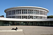
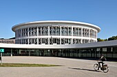
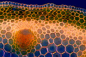
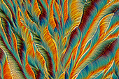
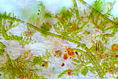
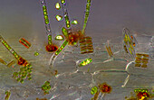
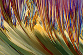
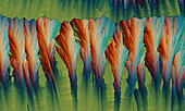
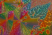
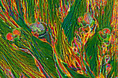
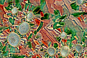
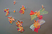
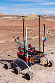
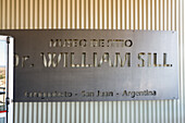
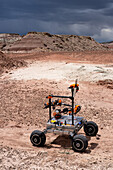
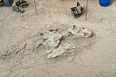
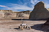
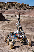
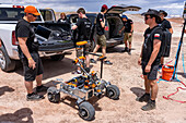
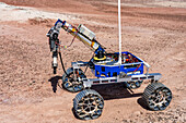
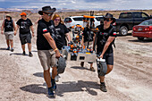
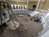
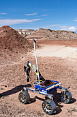
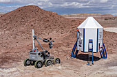
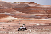
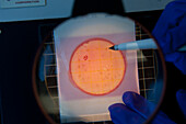
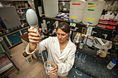
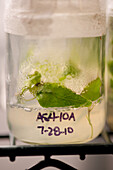
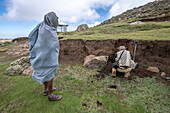
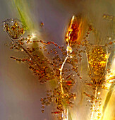
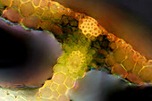
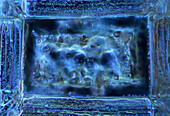
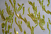
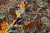
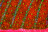
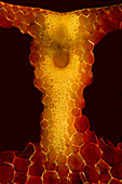
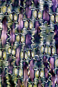
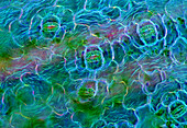
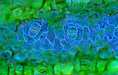
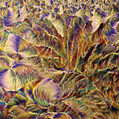
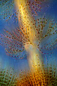
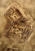
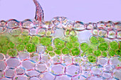
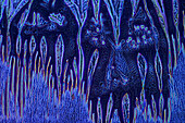
13929309 - France, Nord, Villeneuve d'Ascq, University of Lille, Cité Scientifique campus, circular building housing the library and called LILLIAD (Learning Center Innovation1), a scientific center of the University of Lille (scientific university library, multimedia, large scientific collection, expanded library and digitized resources), student lying in the sun\n13931046 - France, Nord, Villeneuve d'Ascq, University of Lille, Cité Scientifique campus, circular building housing the library and called LILLIAD (Learning Center Innovation1), a scientific center of the University of Lille (scientific university library, multimedia, large scientific collection, expanded library and digitized resources), cycling student\n14129928 - Alexander von Humboldt Memorial in Humboldt University in Berlin Germany13925580 - The image presents Anemone sylvestris stalk in transversal cross-section, photographed through the microscope in polarized light at a magnification of 200X\n13925536 - The image presents crystallized paracetamol, photographed through the microscope in polarized light at a magnification of 100X\n13925533 - The image presents crystallized ammonium chloride photographed through the microscope in polarized light at a magnification of 100X\n13925525 - The image presents Carex sp. leaf in transversal cross-section, photographed through the microscope in polarized light at a magnification of 100X\n13925473 - The image presents stomata in Spathiphyllum leaf epidermis, photographed through the microscope in polarized light at a magnification of 100X\n13925385 - The image presents Fragilaria sp., a kind of diatoms against Batrachospermum, a kind of red algae, photographed through the microscope in polarized light at a magnification of 200X\n13925332 - The image presents stomata in hosta leaf epidermis, photographed through the microscope in polarized light at a magnification of 100X\n13925278 - The image presents various tiny algae settled on Lemna sp. root, photographed through the microscope in polarized light at a magnification of 400X. On the right are visible diatoms closed in a special protecting case.\n13925207 - The image presents stomata in Spathiphyllum leaf epidermis, photographed through the microscope in polarized light at a magnification of 400X\n13925161 - The image presents red wine photographed through the microscope in polarized light at a magnification of 100X\n13925148 - The image presents stomata in Spathiphyllum leaf epidermis, photographed through the microscope in polarized light at a magnification of 400X\n13925117 - The image presents crystallized soy sauce, photographed through the microscope in polarized light at a magnification of 100X\n13925085 - The image presents vascular bundles in senecio stalk, photographed through the microscope in polarized light at a magnification of 200X\n13925083 - The image presents Utricularia trap, a kind of carnivorous plant, photographed through the microscope in polarized light and dark field, at a magnification of 100X\n13925079 - The image presents crystallized mixture of kitchen salt and erythritol, photographed through the microscope in polarized light at a magnification of 100X\n13925044 - The image presents various tiny algae settled on Lemna sp. root, photographed through the microscope in polarized light at a magnification of 200X\n13925041 - The image presents various tiny algae settled on Lemna sp. root, photographed through the microscope in polarized light at a magnification of 200X\n13925035 - The image presents crystallized mixture of kitchen salt and erythritol, photographed through the microscope in polarized light at a magnification of 100X\n13925027 - The image presents crystallized soy sauce, photographed through the microscope in polarized light at a magnification of 100X\n13925010 - The image presents read leaf in transversal cross-section, photographed through the microscope in polarized light at a magnification of 100X\n13924998 - The image presents crystallized resorcinol, photographed through the microscope in polarized light at a magnification of 100X\n13924862 - The image presents a single crystal of recrystallized kitchen salt, photographed through the microscope in polarized light at a magnification of 200X\n13924844 - The image presents crystallized mixture of urea and paracetamol, photographed through the microscope in polarized light at a magnification of 100X\n13924827 - The image presents crystallized soy sauce, photographed through the microscope in polarized light at a magnification of 100X\n13924810 - The image presents crystallized soy sauce, photographed through the microscope in polarized light at a magnification of 100X\n13924754 - The image presents crystallized mixture of malic acid and hydroquinone photographed through the microscope in polarized light at a magnification of 100X\n13924619 - The image presents crystallized tartaric acid, photographed through the microscope in polarized light at a magnification of 100X\n13924584 - The image presents crystallized soy sauce, photographed through the microscope in polarized light at a magnification of 100X\n13924550 - The image presents a single crystal of recrystallized kitchen salt, photographed through the microscope in polarized light at a magnification of 200X\n13924465 - The image presents crystallized silver nitrate, photographed through the microscope in polarized light at a magnification of 100X\n13924437 - The image presents crystallized sulfur, photographed through the microscope in polarized light at a magnification of 100X\n13924395 - The image presents crystallized tartaric acid, photographed through the microscope in polarized light at a magnification of 100X\n13924337 - The image presents tissues in nettle stalk in longitudinal cross section, photographed through the microscope in polarized light at a magnification of 100X\n13924325 - The image presents crystallized mixture of malic acid, salicylic acid and acetanilid, photographed through the microscope in polarized light at a magnification of 100X\n13900338 - Northeastern University Mars Rover. University Rover Challenge, Mars Desert Research Station, Utah. Northeastern University Mars Rover Team, Boston, USA13900314 - Sign on the Dr. William Sill Site Museum in Ischigualasto Provincial Park in San Juan Province, Argentina.13900105 - Mars Rover of the Project Scorpio Team. University Rover Challenge, Mars Desert Research Station, Utah. Wroclaw University of Science and Technology, Poland.13900030 - Actual dinosaur bones in a reconstuction of a dinosaur dig camp in the William Sill Museum in Ischigualasto Provincial Park, Argentina.13899613 - The PCZ Mars Rover in the University Rover Challenge, Mars Desert Research Station in the Mars-like desert in Utah. PCZ Rover Team, Czestochowa University of Technology, Poland13899532 - The PCZ Mars Rover in the University Rover Challenge, Mars Desert Research Station in the Mars-like desert in Utah. PCZ Rover Team, Czestochowa University of Technology, Poland13899351 - Mars Rover of the Project Scorpio Team. University Rover Challenge, Mars Desert Research Station, Utah. Wroclaw University of Science and Technology, Poland.13899279 - The PCZ Mars Rover in the University Rover Challenge, Mars Desert Research Station in the Mars-like desert in Utah. PCZ Rover Team, Czestochowa University of Technology, Poland13899164 - The OzU Mars Rover in the University Rover Challenge, Mars Desert Research Station in the Mars-like desert in Utah. Ozyegin University, Istanbul, Turkey13899060 - Mars Rover of the Project Scorpio Team. University Rover Challenge, Mars Desert Research Station, Utah. Wroclaw University of Science and Technology, Poland.13898698 - A reconstuction of a dinosaur dig camp in the William Sill Museum in Ischigualasto Provincial Park, San Juan, Argentina. Actual dinosaur bones are displayed where they have been partially excavated.13898269 - The OzU Mars Rover in the University Rover Challenge, Mars Desert Research Station in the Mars-like desert in Utah. Ozyegin University, Istanbul, Turkey13897631 - The Binghamton University Mars Rover approaches the Mars Lander in the University Rover Challenge. Mars Desert Research Station, Utah.13897612 - Mars Rover of the RoverOva Team. University Rover Challenge, Mars Desert Research Station, Utah. RoverOva, VSB - Technical University of Ostrava, Czech Republic.13848211 - Woman conducting research in an aquaculture lab at Delaware State University13847649 - Two men examine beaker together while in lab, Beltsville, MD13847142 - Scientist counting cultures on a petri dish13847106 - Grad student working in science lab13846601 - Research testing on poplar trees13846345 - Local Ethiopian farmers look on as a field researcher examines the soil of a hillside, Debre Berhan, Ethiopia.13846342 - Scientist in laboratory with purple liquid13846325 - Scientist conducting experiment in lab13846313 - Scientist inspects captured insects in a tube of liquid13846088 - Scientist doing research in lab13845061 - Close up look at female scientist analyzing petri dish containing samples of peanut plant roots, Tifton, Georgia.13844672 - Dissection of a Brain in science lab13777063 - Benjamin Franklin statue in a garden with trees and blossoming plants; Paris, France13925579 - The image presents stomata in Spathiphyllum leaf epidermis, photographed through the microscope in polarized light at a magnification of 200X\n13925520 - The image presents crystallized callus remover, photographed through the microscope in polarized light at a magnification of 100X.\n13925423 - The image presentstwo suctorians ( a kind of ciliate) and tiny diatoms, photographed through the microscope in polarized light at a magnification of 200X\n13925392 - The image presents reed stalk in transversal cross-section, photographed through the microscope in polarized light at a magnification of 200X\n13925377 - The image presents a single crystal of recrystallized salt, photographed through the microscope in polarized light at a magnification of 100X\n13925372 - The image presents a single crystal of recrystallized kitchen salt, photographed through the microscope in polarized light at a magnification of 200X\n13925367 - The image presents Ophrydium sp. ( a kind of colonial ciliates), photographed through the microscope in polarized light at a magnification of 100X\n13925342 - The image presents crystallized malic acid, photographed through the microscope in polarized light at a magnification of 100X\n13925338 - The image presents crystallized soy sauce, photographed through the microscope in polarized light at a magnification of 100X\n13925287 - The image presents tissues in nettle stalk in longitudinal cross-section, photographed through the microscope in polarized light at a magnification of 100X\n13925265 - The image presents nettle tissues in the stalk in longitudinal cross-section, photographed through the microscope in polarized light at a magnification of 100X\n13925174 - The image presents crystallized tartaric acid, photographed through the microscope in polarized light at a magnification of 100X\n13925104 - The image presents diptera eggs, photographed through the microscope in polarized light at a magnification of 100X\n13925100 - The image presents stomata in Stromanthe sp. leaf epidermis, photographed through the microscope in polarized light at a magnification of 100X\n13925093 - The image presents a single vascular bundle in Carex sp. stalk, photographed through the microscope in polarized light and dark field at a magnification of 200X\n13925078 - The image presents stomata in lily leaf epidermis, photographed through the microscope in polarized light at a magnification of 200X\n13925070 - The image presents stomata in Spathiphyllum sp. leaf epidermis, photographed through the microscope in polarized light at a magnification of 100X\n13925038 - The image presents stomata in Spathiphyllum leaf epidermis, photographed through the microscope in polarized light at a magnification of 200X\n13924900 - The image presents stomata in Spathiphyllum sp. leaf epidermis, photographed through the microscope in polarized light at a magnification of 100X\n13924894 - The image presentstwo suctorians ( a kind of ciliate) and tiny diatoms, photographed through the microscope in polarized light at a magnification of 200X\n13924892 - The image presents crystallized mixture of urea, paracetamol and resorcinol, photographed through the microscope in polarized light at a magnification of 100X\n13924873 - The image presents crystallized mixture of malic acid, salicylic acid and acetanilid, photographed through the microscope in polarized light at a magnification of 100X\n13924866 - The image presents reed stalk in transversal cross-section, photographed through the microscope in polarized light at a magnification of 200X\n13924859 - "The image presents Cladophora sp. ""twigs"" (a kind of green algae) with Cocconeis sp. (a kin of diatoms) settled on it, photographed through the microscope in polarized light at a magnification of 200X"\n13924829 - The image presents crystallized mixture of erythritol and TRIS, photographed through the microscope in polarized light at a magnification of 100X\n13924803 - The image presents Fragilaria sp., a kind of diatoms against Batrachospermum, a kind of red algae, photographed through the microscope in polarized light at a magnification of 200X\n13924794 - The image presents crystallized soy sauce, photographed through the microscope in polarized light at a magnification of 100X\n13924786 - The image presents knautia arvensis tissues in the transversal section of the stalk, photographed through the microscope in bright field, at a magnification of 100X\n13924785 - The image presents diatoms (mostly Gomphonema sp.) photographed through the microscope in polarized light at a magnification of 200X\n13924768 - The image presents crystallized mixture os sugar and salt, photographed through the microscope in polarized light at a magnification of 100X\n13924764 - The image presents Batrachospermum sp., a kind of red algae, photographed through the microscope in polarized light at a magnification of 200X\n13924752 - The image presents crystals of recrystallized kitchen salt, photographed through the microscope in polarized light at a magnification of 100X\n13924748 - The image presents a single stoma in Knautia arvensis epidermis, photographed through the microscope in polarized light at a magnification of 200X\n13924747 - The image presents crystallized soy sauce photographed through the microscope in polarized light and phase contrast at a magnification of 100X\n13924612 - The image presents crystals of recrystallized kitchen salt, photographed through the microscope in polarized light at a magnification of 100X\npage suivante