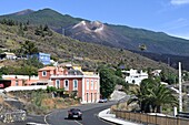
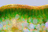
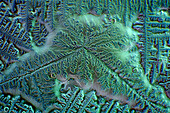
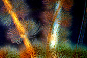
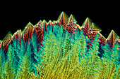
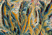
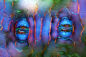
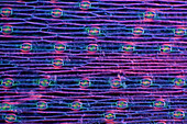
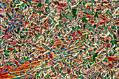
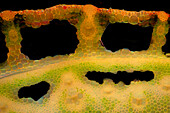
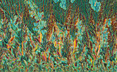
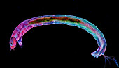
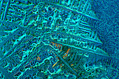
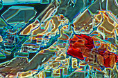
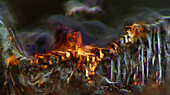
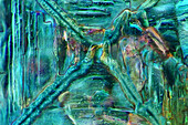
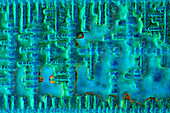
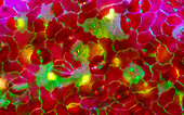
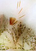
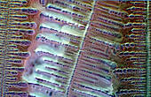
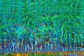
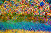
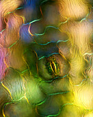
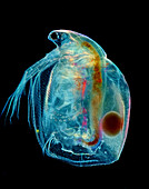
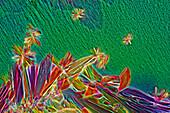
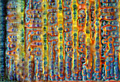
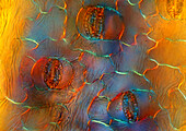
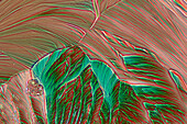
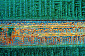
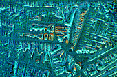
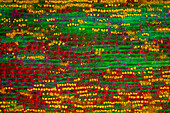
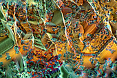
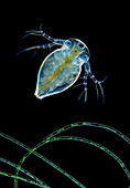
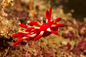
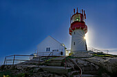
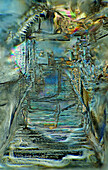
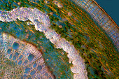
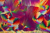
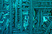
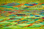
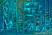
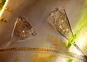
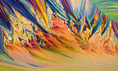
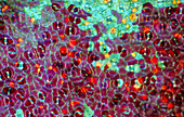
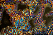
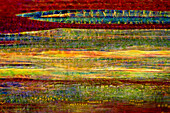
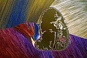
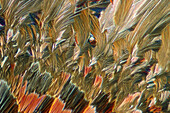
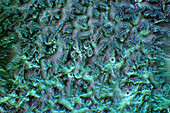
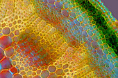
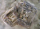
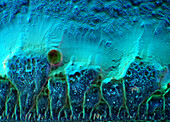
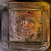
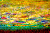
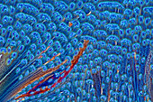
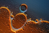
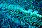
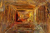
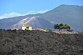
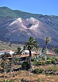
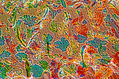
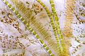
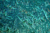
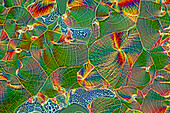
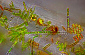
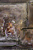
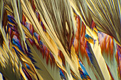
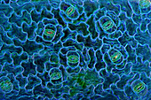
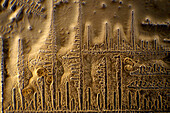
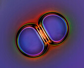
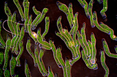
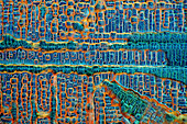
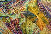
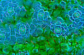
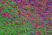
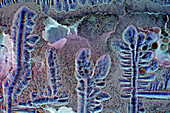
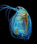
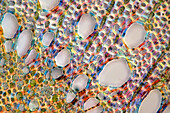
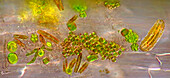
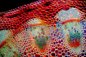
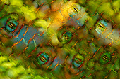
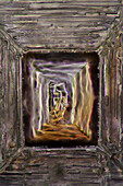
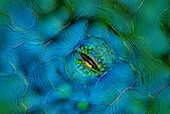
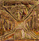
71439589 - New Tajogaite volcano, erupted on September 19th, 2021 for 3 months, photographed in May 2023, west coast of La Palma, Canary Islands, Spain13925468 - The image presents palisade mesophyll in hyacinthus leaf (transversal cross-section) photographed through the microscope in polarized light at a magnification of 200X\n13925453 - The image presents crystallized soy sauce, photographed through the microscope in polarized light at a magnification of 100X\n13925399 - The image presents Batrachospermum sp., a kind of red algae, and FRagilaria sp., a kind of diatoms, photographed through the microscope in polarized light and dark field at a magnification of 100X\n13925388 - The image presents crystallized tartaric acid, photographed through the microscope in polarized light at a magnification of 100X\n13925313 - The image presents crystallized paracetamol, photographed through the microscope in polarized light at a magnification of 100X\n13925303 - The image presents stomata in Spathiphyllum leaf epidermis, photographed through the microscope in polarized light at a magnification of 400X\n13925259 - The image presents stomata in hyacinth leaf epidermis, photographed through the microscope in polarized light at a magnification of 100X\n13925180 - The image presents crystallized tartaric acid, photographed through the microscope in polarized light at a magnification of 100X\n13925106 - The image presents reed stalk in the transversal cross-section, photographed through the microscope in polarized light and dark field, at a magnification of 100X\n13925099 - The image presents crystallized callus remover, photographed through the microscope in polarized light at a magnification of 100X\n13925069 - The image presents diptera larva, photographed through the microscope in polarized light at a magnification of 100X\n13925039 - The image presents crystallized soy sauce, photographed through the microscope in polarized light at a magnification of 100X\n13925022 - The image presents crystallized glycinel, photographed through the microscope in polarized light at a magnification of 100X\n13924981 - The image presents crystallized mixture of kitchen salt and erythritol, photographed through the microscope in polarized light at a magnification of 100X\n13924964 - The image presents crystallized mixture of kitchen salt and erythritol, photographed through the microscope in polarized light at a magnification of 100X\n13924950 - The image presents crystallized soy sauce, photographed through the microscope in polarized light at a magnification of 100X\n13924924 - The image presents stomata in Croton leaf epidermis, photographed through the microscope in polarized light at a magnification of 200X\n13924902 - The image presents Batrachospermum sp., a kind of red algae and some kind of diatoms photographed through the microscope in slightly polarized light at a magnification of 200X\n13924896 - The image presents crystallized soy sauce, photographed through the microscope in polarized light at a magnification of 100X\n13924882 - The image presents crystallized soy sauce, photographed through the microscope in polarized light at a magnification of 100X\n13924809 - The image presents nettle tissues in the transversal cross-section of the stalk, photographed through the microscope in polarized light at a magnification of 200X\n13924776 - The image presents crystallized mixture of kitchen salt and erythritol, photographed through the microscope in polarized light at a magnification of 100X\n13924732 - The image presents a single stoma in Spathiphyllum leaf epidermis, photographed through the microscope in polarized light at a magnification of 200X\n13924713 - The image presents Simocephalus sp. with eggs, a kind of cladoceran, photographed through the microscope in polarized light at a magnification of 100X\n13924640 - The image presents crystallized mixture of myoinositol and tartaric acid, photographed through the microscope in polarized light at a magnification of 100X\n13924620 - The image presents crystallized soy sauce, photographed through the microscope in polarized light at a magnification of 100X\n13924539 - The image presents stomata in Spathiphyllum leaf epidermis, photographed through the microscope in polarized light at a magnification of 200X\n13924524 - The image presents crystallized resorcinol, photographed through the microscope in polarized light at a magnification of 100X\n13924505 - The image presents crystallized soy sauce, photographed through the microscope in polarized light at a magnification of 100X\n13924496 - The image presents crystallized soy sauce, photographed through the microscope in polarized light at a magnification of 100X\n13924490 - The image presents tissues in nettle stalk in longitudinal cross-section, photographed through the microscope in polarized light at a magnification of 100X. The round yellow structures are druses. Druses are the structures created by calcium oxalate.\n13924379 - The image presents mixture of sugar and salt, crystallized photographed through the microscope in polarized light at a magnification of 100X\n13924355 - The image presents crystallized mixture of paracetamol and resorcinol, photographed through the microscope in polarized light at a magnification of 100X\n13924321 - The image presents a single crystal of recrystallized kitchen salt, photographed through the microscope in polarized light at a magnification of 200X\n13924317 - The image presents cladoceran in rare frontal view above filamntous algae photographed through the microscope in polarized light and dark field at a magnification of 100X\n13924309 - The image presents crystallized soy sauce photographed through the microscope in bright field at a magnification of 100X\n13776256 - This nudibranch (Okenia nakamotoensis) is known from the Japan, Indonesia, Philippines and Enewetak and Kwajalein Atolls in the Marshall Islands along with Sipidan Island in Malaysia where this individual was photographed; Malaysia71442909 - Norway, Lindesnes Fyr, lighthouse on the South Cape, most photographed lighthouse in Norway13925626 - The image presents crystallized mixture of kitchen salt and erythritol, photographed through the microscope in polarized light at a magnification of 100X\n13925603 - The image presents oak tissues in transversal cross-section of the stalk, photographed through the microscope in polarized light at a magnification of 100X\n13925567 - The image presents crystallized mixture of urea and paracetamol, photographed through the microscope in polarized light at a magnification of 100X\n13925557 - The image presents crystallized soy sauce, photographed through the microscope in polarized light at a magnification of 100X\n13925506 - The image presents crystallized soy sauce photographed through the microscope in polarized light at a magnification of 100X\n13925491 - The image presents crystallized mixture of paracetamol, urea and resorcinol, photographed through the microscope in polarized light at a magnification of 100X\n13925454 - The image presents nettle tissues in the stalk in longitudinal cross-section, photographed through the microscope at a magnification of 100X\n13925421 - The image presents crystallized soy sauce, photographed through the microscope in polarized light at a magnification of 100X\n13925400 - The image presentstwo suctorians ( a kind of ciliate), photographed through the microscope in polarized light at a magnification of 200X\n13925233 - The image presents crystallized resorcinol photographed through the microscope in polarized light at a magnification of 100X\n13925219 - The image presents crystals of recrystallized kitchen salt, photographed through the microscope in polarized light at a magnification of 100X\n13925162 - The image presents crystallized soy sauce, photographed through the microscope in polarized light at a magnification of 100X\n13925095 - The image presents crystallized soy sauce, photographed through the microscope in polarized light at a magnification of 100X\n13925048 - The image presents stomata in Croton leaf epidermis, photographed through the microscope in polarized light at a magnification of 100X\n13925042 - The image presents crystallized mixture of sugar and salt, photographed through the microscope in polarized light at a magnification of 100X\n13925009 - The image presents nettle stalk longitudinal cross-section photographed through the microscope in polarized light at a magnification of 100X\n13924963 - The image presents crystallized resorcinol, photographed through the microscope in polarized light at a magnification of 100X\n13924960 - The image presents crystallized mixture of eryhtritol and resorcinol, photographed through the microscope in polarized light at a magnification of 100X\n13924918 - The image presents crystallized soy sauce, photographed through the microscope in polarized light at a magnification of 100X\n13924796 - The image presents knautia arvensis tissues in the transversal section of the stalk, photographed through the microscope in bright field, at a magnification of 100X\n13924750 - The image presents two crystals of recrystallized salt, photographed through the microscope in polarized light at a magnification of 100X\n13924724 - The image presents crystallized soy sauce, photographed through the microscope in polarized light at a magnification of 100X\n13924721 - The image presents a single crystal crystallized salt, photographed through the microscope in polarized light at a magnification of 100X\n13924698 - The image presents nettle tissues in the stalk in longitudinal cross-section, photographed through the microscope at a magnification of 100X\n13924572 - The image presents crystallized mixture of erythritol and TRIS, photographed through the microscope in polarized light at a magnification of 100X\n13924471 - The image presents air bubbles formed in foamed milk, photographed through the microscope in polarized light at a magnification of 100X\n13924410 - The image presents crystallized soy sauce, photographed through the microscope in polarized light at a magnification of 100X\n13924370 - The image presents crystallized mixture of kitchen salt and erythritol, photographed through the microscope in polarized light at a magnification of 100X\n71439591 - Lava from the new Tajogaite volcano, erupted on September 19th, 2021 for 3 months, photographed in May 2023 around Todoque/ Las Manchas, west coast of La Palma, Canary Islands, Spain71439588 - New Tajogaite volcano, erupted on September 19th, 2021 for 3 months, photographed in May 2023, west coast of La Palma, Canary Islands, Spain13925537 - The image presents crystallized mixture of tartaric acid, malic acid and acetanilid, photographed through the microscope in polarized light at a magnification of 100X\n13925530 - The image presents various algae (Fragilaria sp., a kind of diatoms, Batrachospermum sp., a kind of red algae and filamentous green algae) photographed through the microscope in slightly polarized light at a magnification of 200X\n13925505 - The image presents crystallized soy sauce, photographed through the microscope in polarized light at a magnification of 100X\n13925480 - The image presents crystallized mixture of urea and malic acid, photographed through the microscope in polarized light at a magnification of 100X\n13925464 - The image presents stomata in Stromanthe sp. leaf epidermis, photographed through the microscope in polarized light at a magnification of 100X\n13925443 - The image presents various tiny algae settled on Lemna sp. root, photographed through the microscope in polarized light at a magnification of 200X\n13925413 - The image presents recrystallized salt, photographed through the microscope in polarized light at a magnification of 100X\n13925412 - The image presents crystallized resorcinol, photographed through the microscope in polarized light at a magnification of 100X\n13925405 - The image presents stomata in Spathiphyllum sp. leaf epidermis, photographed through the microscope in polarized light at a magnification of 100X\n13925402 - The image presents crystallized soy sauce, photographed through the microscope in polarized light at a magnification of 100X\n13925390 - The image presents crystallized soy sauce, photographed through the microscope in polarized light at a magnification of 100X\n13925384 - The image presents air bubbles photographed through the microscope in polarized light at a magnification of 100X\n13925361 - The image presents Ophrydium sp. ( a kind of colonial ciliates), photographed through the microscope in polarized light and dark field at a magnification of 100X\n13925290 - The image presents crystallized soy sauce, photographed through the microscope in polarized light at a magnification of 100X\n13925277 - The image presents crystallized sulfur and hydroquinone, photographed through the microscope in polarized light at a magnification of 100X\n13925240 - The image presents stomata in Spathiphyllum leaf epidermis, photographed through the microscope in polarized light at a magnification of 200X\n13925206 - The image presents crystallized mixture of sulfur and erythritol, photographed through the microscope in polarized light at a magnification of 100X\n13925169 - The image presents crystallized soy sauce photographed through the microscope in polarized light at a magnification of 100X\n13925165 - The image presents crystallized mixture of urea and resorcinol, photographed through the microscope in polarized light at a magnification of 100X\n13925163 - The image presents Simocephalus sp., a kind of cladoceran, photographed through the microscope in polarized light at a magnification of 100X\n13925098 - The image presents crystallized mixture of urea and paracetamol, photographed through the microscope in polarized light at a magnification of 100X\n13925092 - The image presents oak xylem tissue in the transversal cross-section of the stalk, photographed through the microscope in polarized light at a magnification of 400X\n13924948 - The image presents various tiny algae settled on Lemna sp. root, photographed through the microscope in polarized light at a magnification of 400X. On the right are visible diatoms closed in a special protecting case.\n13924921 - The image presents senecio tissues in the transversal cross-section through the stalk, photographed through the microscope in polarized light and dark field at a magnification of 100X\n13924913 - The image presents stomata in Spathiphyllum leaf epidermis, photographed through the microscope in polarized light at a magnification of 200X\n13924824 - The image presents a single crystal of recrystallized salt, photographed through the microscope in polarized light at a magnification of 100X\n13924733 - The image presents crystallized soy sauce, photographed through the microscope in bright field at a magnification of 100X\n13924624 - The image presents a single stoma in Spathiphyllum sp. leaf epidermis, photographed through the microscope in polarized light at a magnification of 200X\n13924596 - The image presents crystallized mixture of kitchen salt and erythritol, photographed through the microscope in polarized light at a magnification of 100X\n13924564 - The image presents stomata in Spathiphyllum leaf epidermis, photographed through the microscope in polarized light at a magnification of 200X\npage suivante