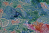
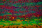
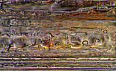
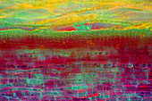
13925576 - The image presents crystallized mixture of erythritol and TRIS, photographed through the microscope in polarized light at a magnification of 100X\n13925553 - The image presents crystallized mixture of sugar and salt, photographed through the microscope in polarized light at a magnification of 100X\n13925552 - The image presents crystallized soy sauce, photographed through the microscope in polarized light at a magnification of 100X\n13925549 - The image presents a small part of forsythia stalk tissues in transversal cross-section, photographed through the microscope in polarized light at a magnification of 100X\n13925548 - The image presents a single crystal of recrystallized kitchen salt, photographed through the microscope in polarized light at a magnification of 200X\n13925446 - The image presents crystals of recrystallized kitchen salt, photographed through the microscope in polarized light at a magnification of 100X\n13925439 - The image presents crystallized soy sauce, photographed through the microscope in polarized light at a magnification of 100X\n13925436 - The image presents crystallized mixture of erythritol and TRIS, photographed through the microscope in polarized light at a magnification of 100X\n13925404 - The image presents crystallized soy sauce, photographed through the microscope in polarized light at a magnification of 100X\n13925394 - The image presents crystallized mixture of salt and sugar, photographed through the microscope in polarized light at a magnification of 100X\n13925359 - The image presents crystallized mixture of kitchen salt and erythritol, photographed through the microscope in polarized light at a magnification of 100X\n13925345 - The image presents nettle tissues in the stalk in transversall cross-section, photographed through the microscope in polarized light and dark field at a magnification of 100X.\n13925297 - The image presents stomata in Spathiphyllum leaf epidermis, photographed through the microscope in polarized light at a magnification of 100X\n13925243 - The image presents various tiny algae settled on Lemna sp. root, photographed through the microscope in polarized light at a magnification of 200X\n13925241 - The image presents nettle tisues in the transversal cross-section of the stalk, photographed through the microscope in polarized light at a magnification of 200X\n13925237 - The image presents crystallized mixture of kitchen salt and erythritol, photographed through the microscope in polarized light at a magnification of 100X\n13925205 - The image presents crystallized soy sauce, photographed through the microscope in polarized light at a magnification of 100X\n13925201 - The image presents a single crystal of recrystallized salt, photographed through the microscope in polarized light at a magnification of 100X\n13925164 - The image presents crystallized mixture of tartaric acid and TRIS, photographed through the microscope in polarized light at a magnification of 100X\n13924997 - The image presents diatoms closed in a special kind of the protecting case, photographed through the microscope in polarized light at a magnification of 400X\n13924994 - The image presents crystallized mixture of erythritol and paracetamol, photographed through the microscope in polarized light at a magnification of 100X\n13924986 - The image presents crystallized tartaric acid, photographed through the microscope in polarized light at a magnification of 100X\n13924965 - The image presents crystallized copper acetate photographed through the microscope in polarized light at a magnification of 100X\n13924884 - The image presents a single crystal of recrystallized kitchen salt, photographed through the microscope in polarized light at a magnification of 200X\n13924875 - The image presents crystallized mixture of erythritol, hydroquinone and TRIS, photographed through the microscope in polarized light at a magnification of 100X\n13924841 - The image presents tissues in nettle stalk in longitudinal cross-section, photographed through the microscope in polarized light at a magnification of 100X. The round yellow structures on the bottom are druses. Druses are the structures created by calcium oxalat.\n13924691 - The image presents recrystallized crystal of salt, photographed through the microscope in polarized light at a magnification of 100X\n13924686 - The image presents nettle tissues in longitudinal cross-section of the stalk, photographed through the microscope in polarized light at a magnification of 100X. Small, round orange particles are the crystals of calcium oxalate called druses.\n13924655 - The image presents stomata in lily leaf epidermis, photographed through the microscope in polarized light at a magnification of 200X\n13924617 - The image presents crystallized callus remover, photographed through the microscope in polarized light at a magnification of 100X\n13924592 - The image presents crystallized soy sauce, photographed through the microscope in polarized light at a magnification of 100X\n13924580 - The image presents reed stalk in transversal cross-section, photographed through the microscope in bright field, at a magnification of 100X\n13924544 - The image presents Batrachospermum sp., a kind of red algae and Fragilaria sp., a kind of diatoms, photographed through the microscope in polarized light at a magnification of 200X\n13924508 - The image presents crystallized soy sauce, photographed through the microscope in polarized light at a magnification of 100X\n13924485 - The image presents crystallized mixture of kitchen salt and erythritol, photographed through the microscope in polarized light at a magnification of 100X\n13924473 - The image presents crystallized callus remover, photographed through the microscope in polarized light at a magnification of 100X\n13924378 - The image presents crystallized mixture of urea paracetamol and resorcinol, photographed through the microscope in polarized light at a magnification of 100X\n13924351 - The image presents crystallized mixture of urea and resorcinol, photographed through the microscope in polarized light at a magnification of 100X\n13924333 - The image presents crystallized soy sauce photographed through the microscope in polarized light at a magnification of 100X\n13924313 - The image presents crystallized mixture of urea and mailc acid, photographed through the microscope in polarized light at a magnification of 100X\n70059649 - Microscope70087670 - A woman, graduand, working on an Atomic force microscope, Organic Semiconductor on the monitor, Clean Room, Nanosystems, LMU, Faculty for Physics, LMU, University, Ludwig Maximilians Universität, Munich, Bavaria, Germany70277695 - children investigate with a microscope in the Springe Museum, region Hannover, Lower Saxony, northern Germany70487390 - 'A real snowflake showing the classic 6-sided star shape, photographed under a microscope;Anchorage, alaska, united states of america'70487387 - 'A real snowflake showing the classic 6-sided star shape, photographed under a microscope;Anchorage, alaska, united states of america'70487388 - 'A real snowflake showing the classic 6-sided star shape, photographed under a microscope;Anchorage, alaska, united states of america'70487386 - 'A real snowflake showing the classic 6-sided star shape, photographed under a microscope;Anchorage, alaska, united states of america'70487389 - 'A real snowflake showing the classic 6-sided star shape, photographed under a microscope;Anchorage, alaska, united states of america'70438622 - Scientist using microscope in lab70474623 - A light microscope examining a sample of tissue in lab for pharmaceutical research70474621 - Female microbiologist viewing specimen under microscope in lab70336814 - Biotitic granite Igneous rocks Pyrenees Spain Petrograhic microscope70059648 - Microscope70474624 - Blood testing, A light microscope examining a sample of blood in a lab70336819 - Slate Metamorphic rock Pyrenees Spain Petrograhic microscope70336817 - Basalt Igneous rocks Petrographic microscope70336816 - Biotitic granite Igneous rocks Pyrenees Spain Petrograhic microscope70336812 - Basalt Igneous rocks Petrographic microscope70474625 - A light microscope examining a sample in lab for pharmaceutical research70474620 - Female microbiologist viewing specimen under microscope in lab70336818 - Quartzite Metamorphic rocks Tarragona Spain Petrographic microscope70336815 - Gneiss rock Metamorphic rock The Costa Brava Gerona Spain Petrographic microscope70336811 - Slate Metamorphic rock Pyrenees Spain Petrograhic microscope70474626 - A light microscope examining a sample in lab for pharmaceutical research70509036 - A lab technician looking into a microscope in a laboratory70509027 - A lab technician looking into a microscope in a laboratory70479470 - Senior Caucasian scientist using microscope in lab, Cape Town, Western Cape, South Africa71086353 - Mixed race scientist using microscope70465922 - African scientist peering into microscope, New Jersey70508573 - Boy looking through microscope, teacher watching70509033 - A lab technician looking into a microscope in a laboratory70509024 - A lab technician using a microscope, looking at camera70508569 - Boy using microscope, close-up13848603 - Microscope10244070 - Woman looking at paintings through microscope in The Museum of Modern Art, New York, USA70336874 - Red Blood Cells Scanning electron microscope71191562 - Cropped image of scientist holding microscope slide over box at laboratory13846313 - Scientist inspects captured insects in a tube of liquid13845669 - Male scientist in lab using pipette surrounded by lab equipment.13848440 - GMO plant tissue on a Petri Dish13846242 - Male scientist in lab using pipette.13844698 - Scientist working in lab with plant material13844639 - Microscopic view of mosquito larvae in a Petri dish13845035 - Insects in a petri dish for inspection in laboratory13996909 - Diamond polishing workshop in a village near Dediapada in Narmada district,Gujarat,India,Asia70026949 - In Vitro-Fertilisation, Prof.Wuerfel, Klinik Dr.Kruesmann Muenchen70085135 - Operation team during a neurosurgical procedure, INI Hanover, Lower Saxony, Germany70336827 - Collenchyma Stem of nettle 20x70336826 - Sclerenchyma Stem of clematis Cross section 7x70336795 - Salmonella typhi Bactery of typhoid fever Gram70336773 - Closterium Chlorophyta Algae Optic microscopy70336771 - Tabellaria flocculosa Diatom Seawed Algae Optic microscopy70336767 - Paramecium sp Ciliata Protozoans Optic microscopy70338869 - Extreme closeup of the head of a greenbottle Lucilla sp showing the extended mouthparts and structure of the compound eyes70336896 - Cancer cells70336850 - Dicotyledon root Acedera70336846 - Dicotyledon stem Rannunculus70336844 - Monocotyledon stem Elder70336825 - Phloem and xylem Dicotyledon root Corte transversal de Sarsaparrilla 140xnext page