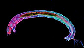
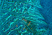
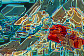
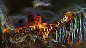
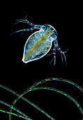
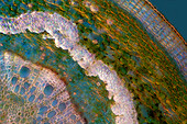
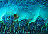
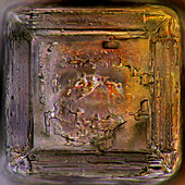
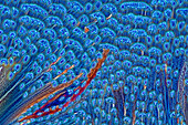
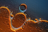
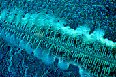
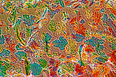
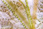
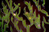
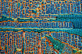
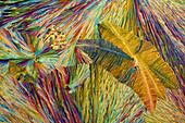
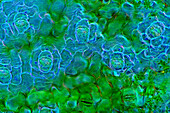
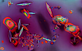
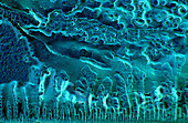
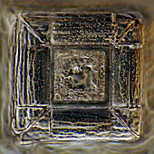
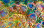
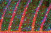
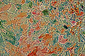
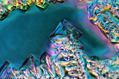
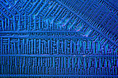
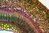
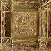
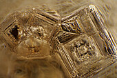
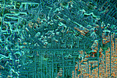
13925069 - The image presents diptera larva, photographed through the microscope in polarized light at a magnification of 100X\n13925039 - The image presents crystallized soy sauce, photographed through the microscope in polarized light at a magnification of 100X\n13925022 - The image presents crystallized glycinel, photographed through the microscope in polarized light at a magnification of 100X\n13924981 - The image presents crystallized mixture of kitchen salt and erythritol, photographed through the microscope in polarized light at a magnification of 100X\n13924964 - The image presents crystallized mixture of kitchen salt and erythritol, photographed through the microscope in polarized light at a magnification of 100X\n13924950 - The image presents crystallized soy sauce, photographed through the microscope in polarized light at a magnification of 100X\n13924924 - The image presents stomata in Croton leaf epidermis, photographed through the microscope in polarized light at a magnification of 200X\n13924902 - The image presents Batrachospermum sp., a kind of red algae and some kind of diatoms photographed through the microscope in slightly polarized light at a magnification of 200X\n13924896 - The image presents crystallized soy sauce, photographed through the microscope in polarized light at a magnification of 100X\n13924882 - The image presents crystallized soy sauce, photographed through the microscope in polarized light at a magnification of 100X\n13924809 - The image presents nettle tissues in the transversal cross-section of the stalk, photographed through the microscope in polarized light at a magnification of 200X\n13924776 - The image presents crystallized mixture of kitchen salt and erythritol, photographed through the microscope in polarized light at a magnification of 100X\n13924732 - The image presents a single stoma in Spathiphyllum leaf epidermis, photographed through the microscope in polarized light at a magnification of 200X\n13924713 - The image presents Simocephalus sp. with eggs, a kind of cladoceran, photographed through the microscope in polarized light at a magnification of 100X\n13924640 - The image presents crystallized mixture of myoinositol and tartaric acid, photographed through the microscope in polarized light at a magnification of 100X\n13924620 - The image presents crystallized soy sauce, photographed through the microscope in polarized light at a magnification of 100X\n13924539 - The image presents stomata in Spathiphyllum leaf epidermis, photographed through the microscope in polarized light at a magnification of 200X\n13924524 - The image presents crystallized resorcinol, photographed through the microscope in polarized light at a magnification of 100X\n13924505 - The image presents crystallized soy sauce, photographed through the microscope in polarized light at a magnification of 100X\n13924496 - The image presents crystallized soy sauce, photographed through the microscope in polarized light at a magnification of 100X\n13924490 - The image presents tissues in nettle stalk in longitudinal cross-section, photographed through the microscope in polarized light at a magnification of 100X. The round yellow structures are druses. Druses are the structures created by calcium oxalate.\n13924379 - The image presents mixture of sugar and salt, crystallized photographed through the microscope in polarized light at a magnification of 100X\n13924355 - The image presents crystallized mixture of paracetamol and resorcinol, photographed through the microscope in polarized light at a magnification of 100X\n13924321 - The image presents a single crystal of recrystallized kitchen salt, photographed through the microscope in polarized light at a magnification of 200X\n13924317 - The image presents cladoceran in rare frontal view above filamntous algae photographed through the microscope in polarized light and dark field at a magnification of 100X\n13925626 - The image presents crystallized mixture of kitchen salt and erythritol, photographed through the microscope in polarized light at a magnification of 100X\n13925603 - The image presents oak tissues in transversal cross-section of the stalk, photographed through the microscope in polarized light at a magnification of 100X\n13925567 - The image presents crystallized mixture of urea and paracetamol, photographed through the microscope in polarized light at a magnification of 100X\n13925557 - The image presents crystallized soy sauce, photographed through the microscope in polarized light at a magnification of 100X\n13925506 - The image presents crystallized soy sauce photographed through the microscope in polarized light at a magnification of 100X\n13925491 - The image presents crystallized mixture of paracetamol, urea and resorcinol, photographed through the microscope in polarized light at a magnification of 100X\n13925421 - The image presents crystallized soy sauce, photographed through the microscope in polarized light at a magnification of 100X\n13925400 - The image presentstwo suctorians ( a kind of ciliate), photographed through the microscope in polarized light at a magnification of 200X\n13925233 - The image presents crystallized resorcinol photographed through the microscope in polarized light at a magnification of 100X\n13925219 - The image presents crystals of recrystallized kitchen salt, photographed through the microscope in polarized light at a magnification of 100X\n13925162 - The image presents crystallized soy sauce, photographed through the microscope in polarized light at a magnification of 100X\n13925095 - The image presents crystallized soy sauce, photographed through the microscope in polarized light at a magnification of 100X\n13925048 - The image presents stomata in Croton leaf epidermis, photographed through the microscope in polarized light at a magnification of 100X\n13925042 - The image presents crystallized mixture of sugar and salt, photographed through the microscope in polarized light at a magnification of 100X\n13925009 - The image presents nettle stalk longitudinal cross-section photographed through the microscope in polarized light at a magnification of 100X\n13924963 - The image presents crystallized resorcinol, photographed through the microscope in polarized light at a magnification of 100X\n13924960 - The image presents crystallized mixture of eryhtritol and resorcinol, photographed through the microscope in polarized light at a magnification of 100X\n13924918 - The image presents crystallized soy sauce, photographed through the microscope in polarized light at a magnification of 100X\n13924750 - The image presents two crystals of recrystallized salt, photographed through the microscope in polarized light at a magnification of 100X\n13924724 - The image presents crystallized soy sauce, photographed through the microscope in polarized light at a magnification of 100X\n13924721 - The image presents a single crystal crystallized salt, photographed through the microscope in polarized light at a magnification of 100X\n13924572 - The image presents crystallized mixture of erythritol and TRIS, photographed through the microscope in polarized light at a magnification of 100X\n13924471 - The image presents air bubbles formed in foamed milk, photographed through the microscope in polarized light at a magnification of 100X\n13924410 - The image presents crystallized soy sauce, photographed through the microscope in polarized light at a magnification of 100X\n13924370 - The image presents crystallized mixture of kitchen salt and erythritol, photographed through the microscope in polarized light at a magnification of 100X\n13925537 - The image presents crystallized mixture of tartaric acid, malic acid and acetanilid, photographed through the microscope in polarized light at a magnification of 100X\n13925530 - The image presents various algae (Fragilaria sp., a kind of diatoms, Batrachospermum sp., a kind of red algae and filamentous green algae) photographed through the microscope in slightly polarized light at a magnification of 200X\n13925505 - The image presents crystallized soy sauce, photographed through the microscope in polarized light at a magnification of 100X\n13925480 - The image presents crystallized mixture of urea and malic acid, photographed through the microscope in polarized light at a magnification of 100X\n13925464 - The image presents stomata in Stromanthe sp. leaf epidermis, photographed through the microscope in polarized light at a magnification of 100X\n13925443 - The image presents various tiny algae settled on Lemna sp. root, photographed through the microscope in polarized light at a magnification of 200X\n13925413 - The image presents recrystallized salt, photographed through the microscope in polarized light at a magnification of 100X\n13925412 - The image presents crystallized resorcinol, photographed through the microscope in polarized light at a magnification of 100X\n13925405 - The image presents stomata in Spathiphyllum sp. leaf epidermis, photographed through the microscope in polarized light at a magnification of 100X\n13925402 - The image presents crystallized soy sauce, photographed through the microscope in polarized light at a magnification of 100X\n13925390 - The image presents crystallized soy sauce, photographed through the microscope in polarized light at a magnification of 100X\n13925384 - The image presents air bubbles photographed through the microscope in polarized light at a magnification of 100X\n13925361 - The image presents Ophrydium sp. ( a kind of colonial ciliates), photographed through the microscope in polarized light and dark field at a magnification of 100X\n13925290 - The image presents crystallized soy sauce, photographed through the microscope in polarized light at a magnification of 100X\n13925277 - The image presents crystallized sulfur and hydroquinone, photographed through the microscope in polarized light at a magnification of 100X\n13925240 - The image presents stomata in Spathiphyllum leaf epidermis, photographed through the microscope in polarized light at a magnification of 200X\n13925206 - The image presents crystallized mixture of sulfur and erythritol, photographed through the microscope in polarized light at a magnification of 100X\n13925169 - The image presents crystallized soy sauce photographed through the microscope in polarized light at a magnification of 100X\n13925165 - The image presents crystallized mixture of urea and resorcinol, photographed through the microscope in polarized light at a magnification of 100X\n13925163 - The image presents Simocephalus sp., a kind of cladoceran, photographed through the microscope in polarized light at a magnification of 100X\n13925098 - The image presents crystallized mixture of urea and paracetamol, photographed through the microscope in polarized light at a magnification of 100X\n13925092 - The image presents oak xylem tissue in the transversal cross-section of the stalk, photographed through the microscope in polarized light at a magnification of 400X\n13924948 - The image presents various tiny algae settled on Lemna sp. root, photographed through the microscope in polarized light at a magnification of 400X. On the right are visible diatoms closed in a special protecting case.\n13924921 - The image presents senecio tissues in the transversal cross-section through the stalk, photographed through the microscope in polarized light and dark field at a magnification of 100X\n13924913 - The image presents stomata in Spathiphyllum leaf epidermis, photographed through the microscope in polarized light at a magnification of 200X\n13924824 - The image presents a single crystal of recrystallized salt, photographed through the microscope in polarized light at a magnification of 100X\n13924624 - The image presents a single stoma in Spathiphyllum sp. leaf epidermis, photographed through the microscope in polarized light at a magnification of 200X\n13924596 - The image presents crystallized mixture of kitchen salt and erythritol, photographed through the microscope in polarized light at a magnification of 100X\n13924564 - The image presents stomata in Spathiphyllum leaf epidermis, photographed through the microscope in polarized light at a magnification of 200X\n13924504 - "The image presents Cladophora sp. ""twigs"" (a kind of green algae) with Cocconeis sp. (a kin of diatoms) settled on it, photographed through the microscope in polarized light at a magnification of 100X"\n13924460 - The image presents crystallized red wine, photographed through the microscope in polarized light at a magnification of 100X\n13924417 - The image presents crystallized soy sauce, photographed through the microscope in polarized light at a magnification of 100X\n13924411 - The image presents a single crystal of recrystallized salt, photographed through the microscope in polarized light at a magnification of 100X\n13924302 - The image presents nettle tissues in the transversal section of the stalk, photographed through the microscope in polarized light at a magnification of 400X\n13925616 - The image presents stomata in Stromanthe sp. leaf epidermis, photographed through the microscope in polarized light at a magnification of 100X\n13925576 - The image presents crystallized mixture of erythritol and TRIS, photographed through the microscope in polarized light at a magnification of 100X\n13925553 - The image presents crystallized mixture of sugar and salt, photographed through the microscope in polarized light at a magnification of 100X\n13925552 - The image presents crystallized soy sauce, photographed through the microscope in polarized light at a magnification of 100X\n13925549 - The image presents a small part of forsythia stalk tissues in transversal cross-section, photographed through the microscope in polarized light at a magnification of 100X\n13925548 - The image presents a single crystal of recrystallized kitchen salt, photographed through the microscope in polarized light at a magnification of 200X\n13925446 - The image presents crystals of recrystallized kitchen salt, photographed through the microscope in polarized light at a magnification of 100X\n13925439 - The image presents crystallized soy sauce, photographed through the microscope in polarized light at a magnification of 100X\n13925436 - The image presents crystallized mixture of erythritol and TRIS, photographed through the microscope in polarized light at a magnification of 100X\n13925404 - The image presents crystallized soy sauce, photographed through the microscope in polarized light at a magnification of 100X\n13925394 - The image presents crystallized mixture of salt and sugar, photographed through the microscope in polarized light at a magnification of 100X\n13925359 - The image presents crystallized mixture of kitchen salt and erythritol, photographed through the microscope in polarized light at a magnification of 100X\n13925345 - The image presents nettle tissues in the stalk in transversall cross-section, photographed through the microscope in polarized light and dark field at a magnification of 100X.\n13925297 - The image presents stomata in Spathiphyllum leaf epidermis, photographed through the microscope in polarized light at a magnification of 100X\n13925243 - The image presents various tiny algae settled on Lemna sp. root, photographed through the microscope in polarized light at a magnification of 200X\nnext page