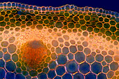
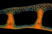
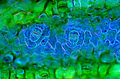
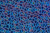
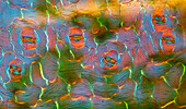
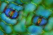
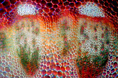
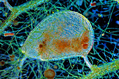
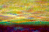
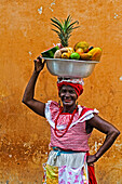
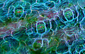
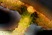
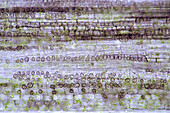
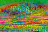
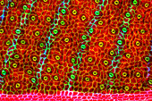
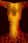
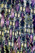
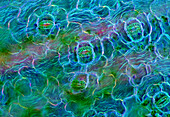
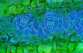
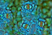
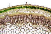
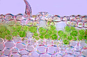
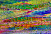
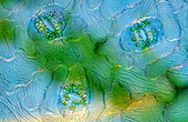
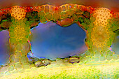
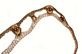
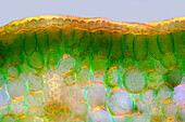
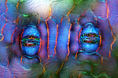
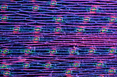
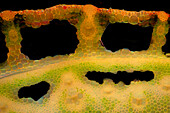
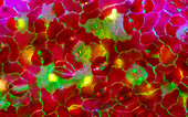
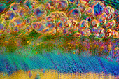
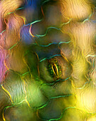
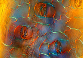
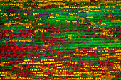
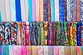
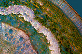
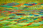
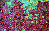
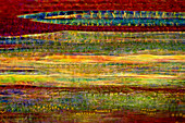
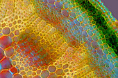
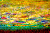
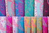
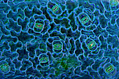
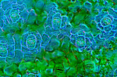
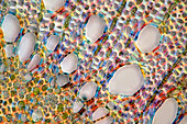
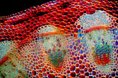
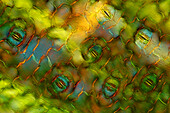
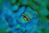
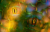
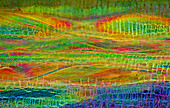
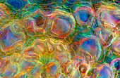
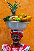
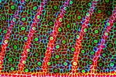
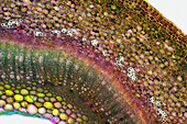
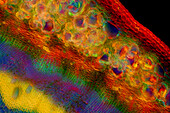
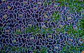
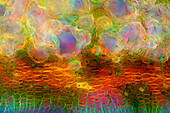
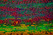
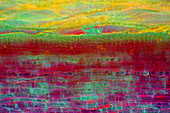
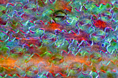
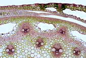
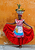
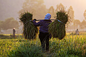
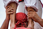
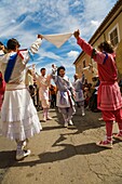
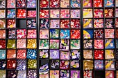
70103060 - yes, Cold, Colds, Color, Colour, Dark-haired, Disease, Diseases, Female, Generation X, Handkerchief70272408 - Butterfly Chrysalids 'Idea Leuconoe' Called The 'Handkerchief', House Of Tropical Butterflies, Naturospace, Honfleur, Calvados (14), Normandy, France13925580 - The image presents Anemone sylvestris stalk in transversal cross-section, photographed through the microscope in polarized light at a magnification of 200X\n13925525 - The image presents Carex sp. leaf in transversal cross-section, photographed through the microscope in polarized light at a magnification of 100X\n13925473 - The image presents stomata in Spathiphyllum leaf epidermis, photographed through the microscope in polarized light at a magnification of 100X\n13925332 - The image presents stomata in hosta leaf epidermis, photographed through the microscope in polarized light at a magnification of 100X\n13925207 - The image presents stomata in Spathiphyllum leaf epidermis, photographed through the microscope in polarized light at a magnification of 400X\n13925148 - The image presents stomata in Spathiphyllum leaf epidermis, photographed through the microscope in polarized light at a magnification of 400X\n13925085 - The image presents vascular bundles in senecio stalk, photographed through the microscope in polarized light at a magnification of 200X\n13925083 - The image presents Utricularia trap, a kind of carnivorous plant, photographed through the microscope in polarized light and dark field, at a magnification of 100X\n13925010 - The image presents read leaf in transversal cross-section, photographed through the microscope in polarized light at a magnification of 100X\n13924337 - The image presents tissues in nettle stalk in longitudinal cross section, photographed through the microscope in polarized light at a magnification of 100X\n13847821 - Palenquera woman sells fruts in Cartagena , Colombia. Palenqueras are a unique African descendat ethnic group found in the north region of South America13925579 - The image presents stomata in Spathiphyllum leaf epidermis, photographed through the microscope in polarized light at a magnification of 200X\n13925392 - The image presents reed stalk in transversal cross-section, photographed through the microscope in polarized light at a magnification of 200X\n13925287 - The image presents tissues in nettle stalk in longitudinal cross-section, photographed through the microscope in polarized light at a magnification of 100X\n13925265 - The image presents nettle tissues in the stalk in longitudinal cross-section, photographed through the microscope in polarized light at a magnification of 100X\n13925100 - The image presents stomata in Stromanthe sp. leaf epidermis, photographed through the microscope in polarized light at a magnification of 100X\n13925093 - The image presents a single vascular bundle in Carex sp. stalk, photographed through the microscope in polarized light and dark field at a magnification of 200X\n13925078 - The image presents stomata in lily leaf epidermis, photographed through the microscope in polarized light at a magnification of 200X\n13925070 - The image presents stomata in Spathiphyllum sp. leaf epidermis, photographed through the microscope in polarized light at a magnification of 100X\n13925038 - The image presents stomata in Spathiphyllum leaf epidermis, photographed through the microscope in polarized light at a magnification of 200X\n13924900 - The image presents stomata in Spathiphyllum sp. leaf epidermis, photographed through the microscope in polarized light at a magnification of 100X\n13924866 - The image presents reed stalk in transversal cross-section, photographed through the microscope in polarized light at a magnification of 200X\n13924786 - The image presents knautia arvensis tissues in the transversal section of the stalk, photographed through the microscope in bright field, at a magnification of 100X\n13924748 - The image presents a single stoma in Knautia arvensis epidermis, photographed through the microscope in polarized light at a magnification of 200X\n13924555 - The image presents tissues in nettle stalk in longitudinal cross-section, photographed through the microscope in polarized light at a magnification of 100X\n13924552 - The image presents stomata in Spathiphyllum leaf epidermis, photographed through the microscope in polarized light at a magnification of 400X\n13924452 - The image presents reed stalk in transversal cross-section, photographed through the microscope in polarized light at a magnification of 200X\n13924434 - The image presents carex sp. leaf in transversal cross-section, photographed through the microscope in polarized light at a magnification of 100X\n13925468 - The image presents palisade mesophyll in hyacinthus leaf (transversal cross-section) photographed through the microscope in polarized light at a magnification of 200X\n13925303 - The image presents stomata in Spathiphyllum leaf epidermis, photographed through the microscope in polarized light at a magnification of 400X\n13925259 - The image presents stomata in hyacinth leaf epidermis, photographed through the microscope in polarized light at a magnification of 100X\n13925106 - The image presents reed stalk in the transversal cross-section, photographed through the microscope in polarized light and dark field, at a magnification of 100X\n13924924 - The image presents stomata in Croton leaf epidermis, photographed through the microscope in polarized light at a magnification of 200X\n13924809 - The image presents nettle tissues in the transversal cross-section of the stalk, photographed through the microscope in polarized light at a magnification of 200X\n13924732 - The image presents a single stoma in Spathiphyllum leaf epidermis, photographed through the microscope in polarized light at a magnification of 200X\n13924539 - The image presents stomata in Spathiphyllum leaf epidermis, photographed through the microscope in polarized light at a magnification of 200X\n13924490 - The image presents tissues in nettle stalk in longitudinal cross-section, photographed through the microscope in polarized light at a magnification of 100X. The round yellow structures are druses. Druses are the structures created by calcium oxalate.\n13826247 - silk scarves, bazaar, Bukhara, Uzbekistan13925603 - The image presents oak tissues in transversal cross-section of the stalk, photographed through the microscope in polarized light at a magnification of 100X\n13925454 - The image presents nettle tissues in the stalk in longitudinal cross-section, photographed through the microscope at a magnification of 100X\n13925048 - The image presents stomata in Croton leaf epidermis, photographed through the microscope in polarized light at a magnification of 100X\n13925009 - The image presents nettle stalk longitudinal cross-section photographed through the microscope in polarized light at a magnification of 100X\n13924796 - The image presents knautia arvensis tissues in the transversal section of the stalk, photographed through the microscope in bright field, at a magnification of 100X\n13924698 - The image presents nettle tissues in the stalk in longitudinal cross-section, photographed through the microscope at a magnification of 100X\n13826246 - silk scarves, bazaar, Bukhara, Uzbekistan13925464 - The image presents stomata in Stromanthe sp. leaf epidermis, photographed through the microscope in polarized light at a magnification of 100X\n13925405 - The image presents stomata in Spathiphyllum sp. leaf epidermis, photographed through the microscope in polarized light at a magnification of 100X\n13925240 - The image presents stomata in Spathiphyllum leaf epidermis, photographed through the microscope in polarized light at a magnification of 200X\n13925092 - The image presents oak xylem tissue in the transversal cross-section of the stalk, photographed through the microscope in polarized light at a magnification of 400X\n13924921 - The image presents senecio tissues in the transversal cross-section through the stalk, photographed through the microscope in polarized light and dark field at a magnification of 100X\n13924913 - The image presents stomata in Spathiphyllum leaf epidermis, photographed through the microscope in polarized light at a magnification of 200X\n13924624 - The image presents a single stoma in Spathiphyllum sp. leaf epidermis, photographed through the microscope in polarized light at a magnification of 200X\n13924564 - The image presents stomata in Spathiphyllum leaf epidermis, photographed through the microscope in polarized light at a magnification of 200X\n13924392 - Tissues in nettle stalk photographed through the microscope\n13924302 - The image presents nettle tissues in the transversal section of the stalk, photographed through the microscope in polarized light at a magnification of 400X\n13847862 - Palenquera woman sells fruts in Cartagena , Colombia. Palenqueras are a unique African descendat ethnic group found in the north region of South America13826092 - Sacred Tree wrapped with scarves as offering, in Wat Chedi Luang temple, Chiang Mai, Thailand13925616 - The image presents stomata in Stromanthe sp. leaf epidermis, photographed through the microscope in polarized light at a magnification of 100X\n13925549 - The image presents a small part of forsythia stalk tissues in transversal cross-section, photographed through the microscope in polarized light at a magnification of 100X\n13925345 - The image presents nettle tissues in the stalk in transversall cross-section, photographed through the microscope in polarized light and dark field at a magnification of 100X.\n13925297 - The image presents stomata in Spathiphyllum leaf epidermis, photographed through the microscope in polarized light at a magnification of 100X\n13925241 - The image presents nettle tisues in the transversal cross-section of the stalk, photographed through the microscope in polarized light at a magnification of 200X\n13924841 - The image presents tissues in nettle stalk in longitudinal cross-section, photographed through the microscope in polarized light at a magnification of 100X. The round yellow structures on the bottom are druses. Druses are the structures created by calcium oxalat.\n13924686 - The image presents nettle tissues in longitudinal cross-section of the stalk, photographed through the microscope in polarized light at a magnification of 100X. Small, round orange particles are the crystals of calcium oxalate called druses.\n13924655 - The image presents stomata in lily leaf epidermis, photographed through the microscope in polarized light at a magnification of 200X\n13924580 - The image presents reed stalk in transversal cross-section, photographed through the microscope in bright field, at a magnification of 100X\n13848297 - Palenquera woman sells fruts in Cartagena , Colombia. Palenqueras are a unique African descendat ethnic group found in the north region of South America71374516 - Canada, Ontario, Boy (8-9) in knit hat lying on sofa covered with blanket70023450 - Pilgrims travelling afoot, on horseback and with oxcarts on the Raya Real, Andalusia, Spain70006542 - Suit, stilllife tie70217764 - Allergic woman70293703 - Travelling by boat from Dubrovnik to Mljet island, Mljet island is in background, Croatia71071627 - Farmer carrying rice plants in rural field70408598 - Xicots de Vilafranca ´Castellers´ building human tower, a Catalan tradition Plaça de la Vila de Gràcia Barri de Gràcia Barcelona, Spain70320703 - Cossiers dance festival, Alaro, Majorca, Balearic Islands, Spain71027847 - Mixed race woman in headscarf outdoors70390432 - Nens del Vendrell ´Castellers´ building human tower, a Catalan tradition Fira de Santa Teresa, town festival Plaça Vella El Vendrell Tarragona province, Catalonia, Spain71335930 - Itsukushima Miyajima Japan. Handkerchiefs70408607 - Xicots de Vilafranca ´Castellers´ building human tower, a Catalan tradition Plaça de la Vila de Gràcia Barri de Gràcia Barcelona, Spain70395118 - young girl at beach looking around71090428 - Caucasian athlete resting after workout70370713 - Close-up of a woman suffering from cold71090432 - Caucasian athlete standing with bicycle71090431 - Caucasian athlete stretching leg71090430 - Caucasian athlete resting after workout70466807 - Mixed race girl at party wearing sunglasses, San Diego, California, United States70465616 - Hispanic woman laughing, Miami, FL71057556 - Dog wearing bandana laying on wooden floor71090433 - Caucasian athlete standing with bicycle71090429 - Caucasian athlete resting after workout71129365 - Caucasian man winding rope70217763 - Allergic woman71185193 - Confident Caucasian woman wearing football jersey71185192 - Close up of Caucasian woman making a face70370158 - Portrait of young woman blowing her nose