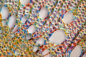
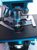
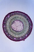
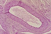
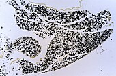
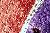
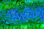
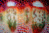
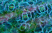
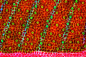
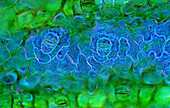
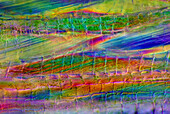
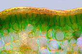
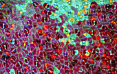
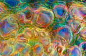
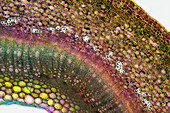
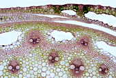
13925092 - The image presents oak xylem tissue in the transversal cross-section of the stalk, photographed through the microscope in polarized light at a magnification of 400X\n13848739 - Plant tissue culture in a test tube13844764 - Plant tissue culture in a test tube13848440 - GMO plant tissue on a Petri Dish70474623 - A light microscope examining a sample of tissue in lab for pharmaceutical research70336876 - Bone tissue Osteon70118922 - Bathroom, Bathrooms, Close up, Close-up, Closeup, Color, Colour, Concept, Concepts, Hygiene, Indoor, Indoors, Inside, Interior, Metal, Object, Objects, Pair, Roll, Rolls, Soft, Still life, Thing, Things, Toilet paper, Toilet tissue, Two, Vertical, White,71139527 - Facelift. Blepharoplasty. Surgery that aims to rejuvenate the look. It is to remove excess skin and bags of both eyelids and periocular tissue remodeling. Plastic surgery. Doctor attending to patient medical consultation.70336892 - Spinal cordon Nervous tissue70336886 - Multipolar neuron of the spinal cordon Nervous tissue 14x70336807 - Section through xylem Vegetable tissue 7x70336797 - Venule Blood vessel Circulatory tissue 35x70336796 - Multipolar neuron of the spinal cordon Nervous tissue 7x70336895 - Epithelial tissue70336890 - Multipolar neuron of the spinal cordon Nervous tissue 140x70336885 - Fat cells Adipose tissue 9x70336870 - Fat cells Adipose tissue70146534 - Bathroom, Bathrooms, Checkered, Chequered, Clean, Color, Colour, Concept, Concepts, Different, Horizontal, Hygiene, Indoor, Indoors, Inside, Interior, Lavatories, Lavatory, Pair, Pipe, Pipes, Restroom, Restrooms, Toilet, Toilet paper, Toilet tissue, Toil71139528 - Facelift. Blepharoplasty. Surgery that aims to rejuvenate the look. It is to remove excess skin and bags of both eyelids and periocular tissue remodeling. Plastic surgery. Doctor attending to patient medical consultation.70480994 - Tissue deposited on the edge of a fountain to allow bees to drink. France.70336867 - Multipolar neuron of the spinal cord Nervous tissue70336799 - Spinal cordon Nervous tissue70336894 - Fat cells Adipose tissue70336893 - Cilia ephitelial tissue70336891 - Electron micrograph of striated muscle tissue70336875 - Cilia ephitelial tissue70336804 - Epithelial tissue of trachea Tracheal epithelium70336801 - Adypocites Adipose tissue 35x70336800 - Conective tissue Mesenteric 140x70336798 - Artery with greasy Blood vessel Circulatory tissue70336887 - Connective tissue 35x70336884 - Connective tissue 35x70336803 - Hyaline cartilage Cartilaginous tissue 9x13925580 - The image presents Anemone sylvestris stalk in transversal cross-section, photographed through the microscope in polarized light at a magnification of 200X\n13925525 - The image presents Carex sp. leaf in transversal cross-section, photographed through the microscope in polarized light at a magnification of 100X\n13925473 - The image presents stomata in Spathiphyllum leaf epidermis, photographed through the microscope in polarized light at a magnification of 100X\n13925332 - The image presents stomata in hosta leaf epidermis, photographed through the microscope in polarized light at a magnification of 100X\n13925207 - The image presents stomata in Spathiphyllum leaf epidermis, photographed through the microscope in polarized light at a magnification of 400X\n13925148 - The image presents stomata in Spathiphyllum leaf epidermis, photographed through the microscope in polarized light at a magnification of 400X\n13925085 - The image presents vascular bundles in senecio stalk, photographed through the microscope in polarized light at a magnification of 200X\n13925083 - The image presents Utricularia trap, a kind of carnivorous plant, photographed through the microscope in polarized light and dark field, at a magnification of 100X\n13925010 - The image presents read leaf in transversal cross-section, photographed through the microscope in polarized light at a magnification of 100X\n13924337 - The image presents tissues in nettle stalk in longitudinal cross section, photographed through the microscope in polarized light at a magnification of 100X\n13847821 - Palenquera woman sells fruts in Cartagena , Colombia. Palenqueras are a unique African descendat ethnic group found in the north region of South America13925579 - The image presents stomata in Spathiphyllum leaf epidermis, photographed through the microscope in polarized light at a magnification of 200X\n13925392 - The image presents reed stalk in transversal cross-section, photographed through the microscope in polarized light at a magnification of 200X\n13925287 - The image presents tissues in nettle stalk in longitudinal cross-section, photographed through the microscope in polarized light at a magnification of 100X\n13925265 - The image presents nettle tissues in the stalk in longitudinal cross-section, photographed through the microscope in polarized light at a magnification of 100X\n13925100 - The image presents stomata in Stromanthe sp. leaf epidermis, photographed through the microscope in polarized light at a magnification of 100X\n13925093 - The image presents a single vascular bundle in Carex sp. stalk, photographed through the microscope in polarized light and dark field at a magnification of 200X\n13925078 - The image presents stomata in lily leaf epidermis, photographed through the microscope in polarized light at a magnification of 200X\n13925070 - The image presents stomata in Spathiphyllum sp. leaf epidermis, photographed through the microscope in polarized light at a magnification of 100X\n13925038 - The image presents stomata in Spathiphyllum leaf epidermis, photographed through the microscope in polarized light at a magnification of 200X\n13924900 - The image presents stomata in Spathiphyllum sp. leaf epidermis, photographed through the microscope in polarized light at a magnification of 100X\n13924866 - The image presents reed stalk in transversal cross-section, photographed through the microscope in polarized light at a magnification of 200X\n13924786 - The image presents knautia arvensis tissues in the transversal section of the stalk, photographed through the microscope in bright field, at a magnification of 100X\n13924748 - The image presents a single stoma in Knautia arvensis epidermis, photographed through the microscope in polarized light at a magnification of 200X\n13924555 - The image presents tissues in nettle stalk in longitudinal cross-section, photographed through the microscope in polarized light at a magnification of 100X\n13924552 - The image presents stomata in Spathiphyllum leaf epidermis, photographed through the microscope in polarized light at a magnification of 400X\n13924452 - The image presents reed stalk in transversal cross-section, photographed through the microscope in polarized light at a magnification of 200X\n13924434 - The image presents carex sp. leaf in transversal cross-section, photographed through the microscope in polarized light at a magnification of 100X\n13925468 - The image presents palisade mesophyll in hyacinthus leaf (transversal cross-section) photographed through the microscope in polarized light at a magnification of 200X\n13925303 - The image presents stomata in Spathiphyllum leaf epidermis, photographed through the microscope in polarized light at a magnification of 400X\n13925259 - The image presents stomata in hyacinth leaf epidermis, photographed through the microscope in polarized light at a magnification of 100X\n13925106 - The image presents reed stalk in the transversal cross-section, photographed through the microscope in polarized light and dark field, at a magnification of 100X\n13924924 - The image presents stomata in Croton leaf epidermis, photographed through the microscope in polarized light at a magnification of 200X\n13924809 - The image presents nettle tissues in the transversal cross-section of the stalk, photographed through the microscope in polarized light at a magnification of 200X\n13924732 - The image presents a single stoma in Spathiphyllum leaf epidermis, photographed through the microscope in polarized light at a magnification of 200X\n13924539 - The image presents stomata in Spathiphyllum leaf epidermis, photographed through the microscope in polarized light at a magnification of 200X\n13924490 - The image presents tissues in nettle stalk in longitudinal cross-section, photographed through the microscope in polarized light at a magnification of 100X. The round yellow structures are druses. Druses are the structures created by calcium oxalate.\n13826247 - silk scarves, bazaar, Bukhara, Uzbekistan13925603 - The image presents oak tissues in transversal cross-section of the stalk, photographed through the microscope in polarized light at a magnification of 100X\n13925454 - The image presents nettle tissues in the stalk in longitudinal cross-section, photographed through the microscope at a magnification of 100X\n13925048 - The image presents stomata in Croton leaf epidermis, photographed through the microscope in polarized light at a magnification of 100X\n13925009 - The image presents nettle stalk longitudinal cross-section photographed through the microscope in polarized light at a magnification of 100X\n13924796 - The image presents knautia arvensis tissues in the transversal section of the stalk, photographed through the microscope in bright field, at a magnification of 100X\n13924698 - The image presents nettle tissues in the stalk in longitudinal cross-section, photographed through the microscope at a magnification of 100X\n13826246 - silk scarves, bazaar, Bukhara, Uzbekistan13925464 - The image presents stomata in Stromanthe sp. leaf epidermis, photographed through the microscope in polarized light at a magnification of 100X\n13925405 - The image presents stomata in Spathiphyllum sp. leaf epidermis, photographed through the microscope in polarized light at a magnification of 100X\n13925240 - The image presents stomata in Spathiphyllum leaf epidermis, photographed through the microscope in polarized light at a magnification of 200X\n13924921 - The image presents senecio tissues in the transversal cross-section through the stalk, photographed through the microscope in polarized light and dark field at a magnification of 100X\n13924913 - The image presents stomata in Spathiphyllum leaf epidermis, photographed through the microscope in polarized light at a magnification of 200X\n13924624 - The image presents a single stoma in Spathiphyllum sp. leaf epidermis, photographed through the microscope in polarized light at a magnification of 200X\n13924564 - The image presents stomata in Spathiphyllum leaf epidermis, photographed through the microscope in polarized light at a magnification of 200X\n13924392 - Tissues in nettle stalk photographed through the microscope\n13924302 - The image presents nettle tissues in the transversal section of the stalk, photographed through the microscope in polarized light at a magnification of 400X\n13847862 - Palenquera woman sells fruts in Cartagena , Colombia. Palenqueras are a unique African descendat ethnic group found in the north region of South America13826092 - Sacred Tree wrapped with scarves as offering, in Wat Chedi Luang temple, Chiang Mai, Thailand13925616 - The image presents stomata in Stromanthe sp. leaf epidermis, photographed through the microscope in polarized light at a magnification of 100X\n13925549 - The image presents a small part of forsythia stalk tissues in transversal cross-section, photographed through the microscope in polarized light at a magnification of 100X\n13925345 - The image presents nettle tissues in the stalk in transversall cross-section, photographed through the microscope in polarized light and dark field at a magnification of 100X.\n13925297 - The image presents stomata in Spathiphyllum leaf epidermis, photographed through the microscope in polarized light at a magnification of 100X\n13925241 - The image presents nettle tisues in the transversal cross-section of the stalk, photographed through the microscope in polarized light at a magnification of 200X\n13924841 - The image presents tissues in nettle stalk in longitudinal cross-section, photographed through the microscope in polarized light at a magnification of 100X. The round yellow structures on the bottom are druses. Druses are the structures created by calcium oxalat.\n13924686 - The image presents nettle tissues in longitudinal cross-section of the stalk, photographed through the microscope in polarized light at a magnification of 100X. Small, round orange particles are the crystals of calcium oxalate called druses.\n13924655 - The image presents stomata in lily leaf epidermis, photographed through the microscope in polarized light at a magnification of 200X\n13924580 - The image presents reed stalk in transversal cross-section, photographed through the microscope in bright field, at a magnification of 100X\n13848297 - Palenquera woman sells fruts in Cartagena , Colombia. Palenqueras are a unique African descendat ethnic group found in the north region of South Americanext page