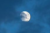
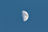
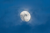
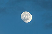
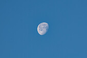
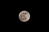
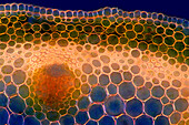
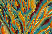
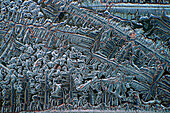
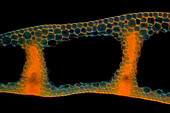
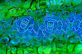
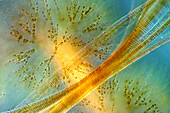
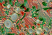
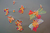
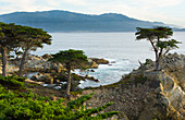
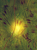
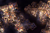
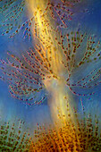
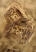
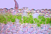
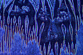
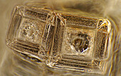
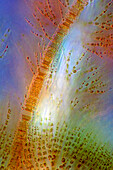
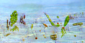
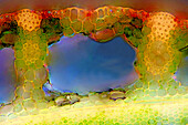
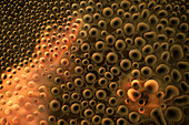
14125134 - A waxing gibbous moon with clouds photographed in the daytime with a telephoto lens over New Mexico.14124082 - A waxing gibbous moon photographed in the daytime with a telephoto lens over New Mexico.14123901 - A waxing crescent moon & stars photographed at night with a telephoto lens over the Dominican Republic.14123763 - A waxing gibbous moon with clouds photographed in the daytime with a telephoto lens over New Mexico.14125824 - A waxing gibbous moon with clouds photographed in the daytime with a telephoto lens over New Mexico.14126163 - A waning gibbous moon photographed in the daytime with a telephoto lens over Utah.14125052 - A full moon photographed with a telephoto lens over Utah.14124005 - A waxing gibbous moon photographed in the daytime with a telephoto lens over Utah.13925580 - The image presents Anemone sylvestris stalk in transversal cross-section, photographed through the microscope in polarized light at a magnification of 200X\n13925536 - The image presents crystallized paracetamol, photographed through the microscope in polarized light at a magnification of 100X\n13925533 - The image presents crystallized ammonium chloride photographed through the microscope in polarized light at a magnification of 100X\n13925525 - The image presents Carex sp. leaf in transversal cross-section, photographed through the microscope in polarized light at a magnification of 100X\n13925473 - The image presents stomata in Spathiphyllum leaf epidermis, photographed through the microscope in polarized light at a magnification of 100X\n13925385 - The image presents Fragilaria sp., a kind of diatoms against Batrachospermum, a kind of red algae, photographed through the microscope in polarized light at a magnification of 200X\n13925332 - The image presents stomata in hosta leaf epidermis, photographed through the microscope in polarized light at a magnification of 100X\n13925278 - The image presents various tiny algae settled on Lemna sp. root, photographed through the microscope in polarized light at a magnification of 400X. On the right are visible diatoms closed in a special protecting case.\n13925207 - The image presents stomata in Spathiphyllum leaf epidermis, photographed through the microscope in polarized light at a magnification of 400X\n13925161 - The image presents red wine photographed through the microscope in polarized light at a magnification of 100X\n13925148 - The image presents stomata in Spathiphyllum leaf epidermis, photographed through the microscope in polarized light at a magnification of 400X\n13925117 - The image presents crystallized soy sauce, photographed through the microscope in polarized light at a magnification of 100X\n13925085 - The image presents vascular bundles in senecio stalk, photographed through the microscope in polarized light at a magnification of 200X\n13925083 - The image presents Utricularia trap, a kind of carnivorous plant, photographed through the microscope in polarized light and dark field, at a magnification of 100X\n13925079 - The image presents crystallized mixture of kitchen salt and erythritol, photographed through the microscope in polarized light at a magnification of 100X\n13925044 - The image presents various tiny algae settled on Lemna sp. root, photographed through the microscope in polarized light at a magnification of 200X\n13925041 - The image presents various tiny algae settled on Lemna sp. root, photographed through the microscope in polarized light at a magnification of 200X\n13925035 - The image presents crystallized mixture of kitchen salt and erythritol, photographed through the microscope in polarized light at a magnification of 100X\n13925027 - The image presents crystallized soy sauce, photographed through the microscope in polarized light at a magnification of 100X\n13925010 - The image presents read leaf in transversal cross-section, photographed through the microscope in polarized light at a magnification of 100X\n13924998 - The image presents crystallized resorcinol, photographed through the microscope in polarized light at a magnification of 100X\n13924862 - The image presents a single crystal of recrystallized kitchen salt, photographed through the microscope in polarized light at a magnification of 200X\n13924844 - The image presents crystallized mixture of urea and paracetamol, photographed through the microscope in polarized light at a magnification of 100X\n13924827 - The image presents crystallized soy sauce, photographed through the microscope in polarized light at a magnification of 100X\n13924810 - The image presents crystallized soy sauce, photographed through the microscope in polarized light at a magnification of 100X\n13924754 - The image presents crystallized mixture of malic acid and hydroquinone photographed through the microscope in polarized light at a magnification of 100X\n13924619 - The image presents crystallized tartaric acid, photographed through the microscope in polarized light at a magnification of 100X\n13924584 - The image presents crystallized soy sauce, photographed through the microscope in polarized light at a magnification of 100X\n13924550 - The image presents a single crystal of recrystallized kitchen salt, photographed through the microscope in polarized light at a magnification of 200X\n13924465 - The image presents crystallized silver nitrate, photographed through the microscope in polarized light at a magnification of 100X\n13924437 - The image presents crystallized sulfur, photographed through the microscope in polarized light at a magnification of 100X\n13924395 - The image presents crystallized tartaric acid, photographed through the microscope in polarized light at a magnification of 100X\n13924337 - The image presents tissues in nettle stalk in longitudinal cross section, photographed through the microscope in polarized light at a magnification of 100X\n13924325 - The image presents crystallized mixture of malic acid, salicylic acid and acetanilid, photographed through the microscope in polarized light at a magnification of 100X\n13823956 - Lucha Libre. After the show. A follower is photographed with cholitas fighters. At left Julieta, in the middle Celia la Simpatica, and Dina , cholitas females wrestlers ,Sports center La Ceja, El Alto, La Paz, Bolivia13810288 - Pebble Beach, California, famous Lone Elm cypress tree and ocean on 17-mile drive one of the most photographed trees in North America71442047 - Rothenburg ob der Tauber, the most photographed Plönlein early in the morning, with Siebersturm, Romantic Road, Bavaria, Germany71439590 - Lava from the new Tajogaite volcano, erupted on September 19th, 2021 for 3 months, photographed in May 2023 around Todoque/ Las Manchas, west coast of La Palma, Canary Islands, Spain13925579 - The image presents stomata in Spathiphyllum leaf epidermis, photographed through the microscope in polarized light at a magnification of 200X\n13925520 - The image presents crystallized callus remover, photographed through the microscope in polarized light at a magnification of 100X.\n13925423 - The image presentstwo suctorians ( a kind of ciliate) and tiny diatoms, photographed through the microscope in polarized light at a magnification of 200X\n13925392 - The image presents reed stalk in transversal cross-section, photographed through the microscope in polarized light at a magnification of 200X\n13925377 - The image presents a single crystal of recrystallized salt, photographed through the microscope in polarized light at a magnification of 100X\n13925372 - The image presents a single crystal of recrystallized kitchen salt, photographed through the microscope in polarized light at a magnification of 200X\n13925367 - The image presents Ophrydium sp. ( a kind of colonial ciliates), photographed through the microscope in polarized light at a magnification of 100X\n13925342 - The image presents crystallized malic acid, photographed through the microscope in polarized light at a magnification of 100X\n13925338 - The image presents crystallized soy sauce, photographed through the microscope in polarized light at a magnification of 100X\n13925287 - The image presents tissues in nettle stalk in longitudinal cross-section, photographed through the microscope in polarized light at a magnification of 100X\n13925265 - The image presents nettle tissues in the stalk in longitudinal cross-section, photographed through the microscope in polarized light at a magnification of 100X\n13925174 - The image presents crystallized tartaric acid, photographed through the microscope in polarized light at a magnification of 100X\n13925104 - The image presents diptera eggs, photographed through the microscope in polarized light at a magnification of 100X\n13925100 - The image presents stomata in Stromanthe sp. leaf epidermis, photographed through the microscope in polarized light at a magnification of 100X\n13925093 - The image presents a single vascular bundle in Carex sp. stalk, photographed through the microscope in polarized light and dark field at a magnification of 200X\n13925078 - The image presents stomata in lily leaf epidermis, photographed through the microscope in polarized light at a magnification of 200X\n13925070 - The image presents stomata in Spathiphyllum sp. leaf epidermis, photographed through the microscope in polarized light at a magnification of 100X\n13925038 - The image presents stomata in Spathiphyllum leaf epidermis, photographed through the microscope in polarized light at a magnification of 200X\n13924900 - The image presents stomata in Spathiphyllum sp. leaf epidermis, photographed through the microscope in polarized light at a magnification of 100X\n13924894 - The image presentstwo suctorians ( a kind of ciliate) and tiny diatoms, photographed through the microscope in polarized light at a magnification of 200X\n13924892 - The image presents crystallized mixture of urea, paracetamol and resorcinol, photographed through the microscope in polarized light at a magnification of 100X\n13924873 - The image presents crystallized mixture of malic acid, salicylic acid and acetanilid, photographed through the microscope in polarized light at a magnification of 100X\n13924866 - The image presents reed stalk in transversal cross-section, photographed through the microscope in polarized light at a magnification of 200X\n13924859 - "The image presents Cladophora sp. ""twigs"" (a kind of green algae) with Cocconeis sp. (a kin of diatoms) settled on it, photographed through the microscope in polarized light at a magnification of 200X"\n13924829 - The image presents crystallized mixture of erythritol and TRIS, photographed through the microscope in polarized light at a magnification of 100X\n13924803 - The image presents Fragilaria sp., a kind of diatoms against Batrachospermum, a kind of red algae, photographed through the microscope in polarized light at a magnification of 200X\n13924794 - The image presents crystallized soy sauce, photographed through the microscope in polarized light at a magnification of 100X\n13924786 - The image presents knautia arvensis tissues in the transversal section of the stalk, photographed through the microscope in bright field, at a magnification of 100X\n13924785 - The image presents diatoms (mostly Gomphonema sp.) photographed through the microscope in polarized light at a magnification of 200X\n13924768 - The image presents crystallized mixture os sugar and salt, photographed through the microscope in polarized light at a magnification of 100X\n13924764 - The image presents Batrachospermum sp., a kind of red algae, photographed through the microscope in polarized light at a magnification of 200X\n13924752 - The image presents crystals of recrystallized kitchen salt, photographed through the microscope in polarized light at a magnification of 100X\n13924748 - The image presents a single stoma in Knautia arvensis epidermis, photographed through the microscope in polarized light at a magnification of 200X\n13924747 - The image presents crystallized soy sauce photographed through the microscope in polarized light and phase contrast at a magnification of 100X\n13924612 - The image presents crystals of recrystallized kitchen salt, photographed through the microscope in polarized light at a magnification of 100X\n13924607 - The image presents crystallized mixture of urea and resorcinol, photographed through the microscope in polarized light at a magnification of 100X\n13924602 - The image presents Fragilaria sp., a kind of diatoms and Batrachospermum sp., a kind of red algae, photographed through the microscope in polarized light at a magnification of 200X\n13924599 - The image presents crystallized soy sauce, photographed through the microscope in polarized light at a magnification of 100X\n13924573 - The image presents air bubbles formed in foemaed milk photographed through the microscope in polarized light at a magnification of 100X\n13924555 - The image presents tissues in nettle stalk in longitudinal cross-section, photographed through the microscope in polarized light at a magnification of 100X\n13924552 - The image presents stomata in Spathiphyllum leaf epidermis, photographed through the microscope in polarized light at a magnification of 400X\n13924513 - The image presents various tiny algae settled on Lemna sp. root, photographed through the microscope in polarized light at a magnification of 200X\n13924489 - The image presents crystallized resorcinol, photographed through the microscope in polarized light at a magnification of 100X\n13924487 - The image presents crystallized mixture of kitchen salt and erythritol, photographed through the microscope in polarized light at a magnification of 100X\n13924452 - The image presents reed stalk in transversal cross-section, photographed through the microscope in polarized light at a magnification of 200X\n13924442 - The image presents tiny air bubbles photographed through the microscope in polarized light at a magnification of 100X\n13924434 - The image presents carex sp. leaf in transversal cross-section, photographed through the microscope in polarized light at a magnification of 100X\n13924422 - The image presents crystallized mixture of kitchen salt and erythritol, photographed through the microscope in polarized light at a magnification of 100X\n13924404 - The image presents euglenoids among green algae, photographed through the microscope in polarized light at a magnification of 100X\n13924398 - The image presents recrystallized sugar photographed through the microscope in polarized light at a magnification of 100X\n13924389 - The image presents recrystallized mixture of salt and erithrytol, photographed through the microscope in polarized light at a magnification of 100X\n13924305 - The image presents crystals of recrystallized kitchen salt, photographed through the microscope in polarized light at a magnification of 100X\n71440993 - Power pole steel construction with many power lines for transporting electricity photographed from directly below against the skynext page