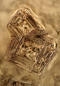
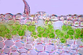
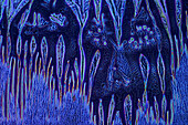
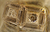
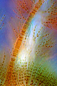
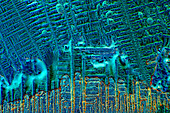
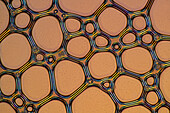
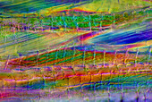
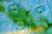
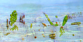
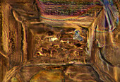
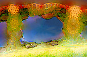
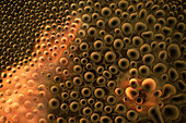
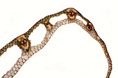
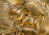
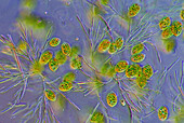
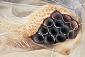
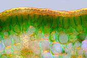
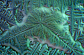
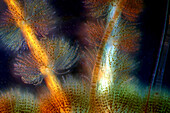
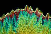
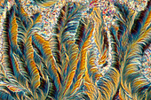
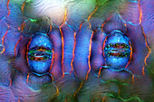
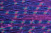
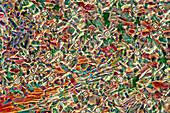
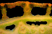
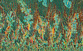
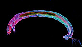
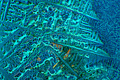
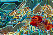
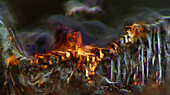
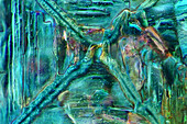
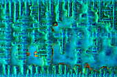
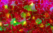
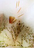
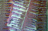
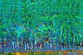
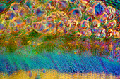
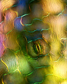
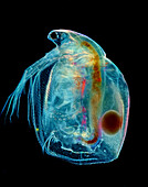
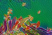
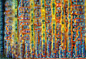
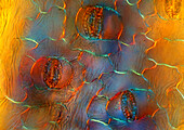
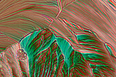
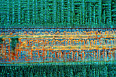
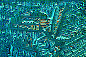
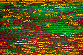
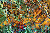
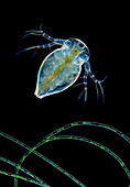
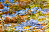
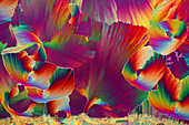
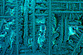
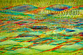
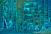
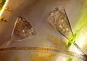
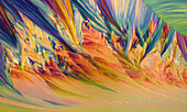
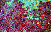
13924752 - The image presents crystals of recrystallized kitchen salt, photographed through the microscope in polarized light at a magnification of 100X\n13924748 - The image presents a single stoma in Knautia arvensis epidermis, photographed through the microscope in polarized light at a magnification of 200X\n13924747 - The image presents crystallized soy sauce photographed through the microscope in polarized light and phase contrast at a magnification of 100X\n13924612 - The image presents crystals of recrystallized kitchen salt, photographed through the microscope in polarized light at a magnification of 100X\n13924607 - The image presents crystallized mixture of urea and resorcinol, photographed through the microscope in polarized light at a magnification of 100X\n13924602 - The image presents Fragilaria sp., a kind of diatoms and Batrachospermum sp., a kind of red algae, photographed through the microscope in polarized light at a magnification of 200X\n13924599 - The image presents crystallized soy sauce, photographed through the microscope in polarized light at a magnification of 100X\n13924573 - The image presents air bubbles formed in foemaed milk photographed through the microscope in polarized light at a magnification of 100X\n13924555 - The image presents tissues in nettle stalk in longitudinal cross-section, photographed through the microscope in polarized light at a magnification of 100X\n13924552 - The image presents stomata in Spathiphyllum leaf epidermis, photographed through the microscope in polarized light at a magnification of 400X\n13924513 - The image presents various tiny algae settled on Lemna sp. root, photographed through the microscope in polarized light at a magnification of 200X\n13924489 - The image presents crystallized resorcinol, photographed through the microscope in polarized light at a magnification of 100X\n13924487 - The image presents crystallized mixture of kitchen salt and erythritol, photographed through the microscope in polarized light at a magnification of 100X\n13924452 - The image presents reed stalk in transversal cross-section, photographed through the microscope in polarized light at a magnification of 200X\n13924442 - The image presents tiny air bubbles photographed through the microscope in polarized light at a magnification of 100X\n13924434 - The image presents carex sp. leaf in transversal cross-section, photographed through the microscope in polarized light at a magnification of 100X\n13924422 - The image presents crystallized mixture of kitchen salt and erythritol, photographed through the microscope in polarized light at a magnification of 100X\n13924404 - The image presents euglenoids among green algae, photographed through the microscope in polarized light at a magnification of 100X\n13924398 - The image presents recrystallized sugar photographed through the microscope in polarized light at a magnification of 100X\n13924389 - The image presents recrystallized mixture of salt and erithrytol, photographed through the microscope in polarized light at a magnification of 100X\n13924305 - The image presents crystals of recrystallized kitchen salt, photographed through the microscope in polarized light at a magnification of 100X\n13900217 - A composite image of the May 9, 2016 transit of Mercury across the Sun, with Mercury at a perfect “inferior conjunction” between the Sun and Earth. Transits happen only rarely, about 13 per century. The next is November 11, 2019. Then in 2032.13899353 - All of Scorpius, plus parts of Lupus and Ara regions of the southern Milky Way. This area was directly overhead when I took this at about 4:30 am local time on April 6, 2014 from near Coonabarabran, Australia. The head of Scorpius is at top his tail at bottom though you could turn this image any direction and it would be correct as seen in the sky at this latitude, depending on the time of night. But in portrait mode like this north is at top. Along the Milky Way are numerous nebulas, including the False Comet area, the Cat's Paw area, and the colourful nebulas around Antares at top. The dark 13898976 - April 17/18, 2001 aurora, taken from home in Alberta. looking south. Part of a series taken looking same direction as substorm hit and subsided, from Image #2 to #15, on Roll #1. (Roll #2 was second camera shooting Provia 100F with 28mm lens and 18mm lens.) All images in this series (#1-02 thru 15) processed in Photoshop with nearly identical enhancements to contrast and colour. Brightness toned down for longer overexposed shots (early ones).13898926 - A single image of bright noctilucent clouds in the dawn sky over a prairie pond near home in southern Alberta on July 7, 2020, with Venus bright as a “morning star” at right above Aldebaran. Comet NEOWISE was in the scene but hidden behind dark weather clouds here.13898452 - A colourful sunrise scene on August 12, 2022, taken from home on the Alberta prairie taken just as the Sun came up. This might be an image useful for sky replacement or a background image.13898238 - An all-sky aurora from Churchill, Manitoba, on Feb 17, 2015, in a frame from a 250-frame time-lapse movie. Taken from the Churchill Northern Studies Centre, using an 8mm Sigma fish-eye lens on the Canon 6D for a 360° view of the sky, though with the camera titled about 25° to create an image suitable for projection in a tilted-dome digital planetarium. This a 15-second exposure at ISO 3200 and f/3.5. The temperature was about -30° C.13897608 - A demo image with the Orion 80mm CF Apo and Celestron AVX mount, with 3 x 8 minute and 3 x 6 minutes, at ISO 1600 with Canon 6D MkII plus shorter 3 x 2 minute and 3 x 1 minute exposures blended in with luminosity masks. Guided with the Orion Starshoot and Orion finderscope, using PHD2, with a lot of wild excursions in the guiding.13897388 - The partial eclipse of the Sun, October 23, 2014, as seen from Jasper, Alberta, shot under clear skies through a mylar filter, on the front of a 66mm f/6 apo refractor using the Canon 60Da for 1/8000 (!) sec exposure at ISO 100. The colours are natural, with the mylar filter providing a neutral “white light” image. The big sunspot on the Sun that day is just disappearing behind the Moon’s limb. The mylar filter gave a white Sun, its natural colour, but I have tinted the Sun’s disk yellow for a more pleasing view that is not just white Sun/black sky.13876833 - Boats mooring in a harbour with the mirror image in the tranquil water; Majorca, Spain13876738 - USA, Tilt Shift Image of Skaters on Pond at Southern End of Central Park; New York City13876736 - Tilt shift lens image - looking up at Sykscrapers with Stars and Stripes flag in Manhattan, New York. USA.13849299 - Very high magnification shot of a silverfish eye; a special technique was used to reveal all the eye surface texture in order to get a SEM like image, but with colour.13820473 - Artistic and abstract winter image71443870 - Village cemetery of the church of Svatého Petra a Pavla with an image of St. Wenceslaus in Albrechtice nad Vltavou in South Bohemia in the Czech Republic71441011 - Irrigation with an irrigation system on a grain field in dry summer as a drone image from above71433504 - Calm image of the North Sea seen from the beach of Ostend, Belgium.13999239 - This wide-field image frames the end stars of the Big Dipper's handle — Mizar at top,and Alkaid at bottom — and to also include in the frame the bright galaxies Messier 101 (at left) and Messier 51 (at lower right,aka the Whirlpool Galaxy). They are small on this image scale but the image serves for a finder chart illustration of the location of these galaxies relative to the Handle. The famous double star Mizar and Alcor is also obvious at top,as is the red giant star 83 Ursa Majoris. The field is 10° x 15°,so wider than binoculars.13999007 - The star cluster Messier 50 in Monoceros the Unicorn,in a wide-field telephoto image simulating the feld of view of binoculars. The Seagull Nebula shows up at bottom. To the left is the cluster NGC 2353.13925468 - The image presents palisade mesophyll in hyacinthus leaf (transversal cross-section) photographed through the microscope in polarized light at a magnification of 200X\n13925453 - The image presents crystallized soy sauce, photographed through the microscope in polarized light at a magnification of 100X\n13925399 - The image presents Batrachospermum sp., a kind of red algae, and FRagilaria sp., a kind of diatoms, photographed through the microscope in polarized light and dark field at a magnification of 100X\n13925388 - The image presents crystallized tartaric acid, photographed through the microscope in polarized light at a magnification of 100X\n13925313 - The image presents crystallized paracetamol, photographed through the microscope in polarized light at a magnification of 100X\n13925303 - The image presents stomata in Spathiphyllum leaf epidermis, photographed through the microscope in polarized light at a magnification of 400X\n13925259 - The image presents stomata in hyacinth leaf epidermis, photographed through the microscope in polarized light at a magnification of 100X\n13925180 - The image presents crystallized tartaric acid, photographed through the microscope in polarized light at a magnification of 100X\n13925106 - The image presents reed stalk in the transversal cross-section, photographed through the microscope in polarized light and dark field, at a magnification of 100X\n13925099 - The image presents crystallized callus remover, photographed through the microscope in polarized light at a magnification of 100X\n13925069 - The image presents diptera larva, photographed through the microscope in polarized light at a magnification of 100X\n13925039 - The image presents crystallized soy sauce, photographed through the microscope in polarized light at a magnification of 100X\n13925022 - The image presents crystallized glycinel, photographed through the microscope in polarized light at a magnification of 100X\n13924981 - The image presents crystallized mixture of kitchen salt and erythritol, photographed through the microscope in polarized light at a magnification of 100X\n13924964 - The image presents crystallized mixture of kitchen salt and erythritol, photographed through the microscope in polarized light at a magnification of 100X\n13924950 - The image presents crystallized soy sauce, photographed through the microscope in polarized light at a magnification of 100X\n13924924 - The image presents stomata in Croton leaf epidermis, photographed through the microscope in polarized light at a magnification of 200X\n13924902 - The image presents Batrachospermum sp., a kind of red algae and some kind of diatoms photographed through the microscope in slightly polarized light at a magnification of 200X\n13924896 - The image presents crystallized soy sauce, photographed through the microscope in polarized light at a magnification of 100X\n13924882 - The image presents crystallized soy sauce, photographed through the microscope in polarized light at a magnification of 100X\n13924809 - The image presents nettle tissues in the transversal cross-section of the stalk, photographed through the microscope in polarized light at a magnification of 200X\n13924776 - The image presents crystallized mixture of kitchen salt and erythritol, photographed through the microscope in polarized light at a magnification of 100X\n13924732 - The image presents a single stoma in Spathiphyllum leaf epidermis, photographed through the microscope in polarized light at a magnification of 200X\n13924713 - The image presents Simocephalus sp. with eggs, a kind of cladoceran, photographed through the microscope in polarized light at a magnification of 100X\n13924640 - The image presents crystallized mixture of myoinositol and tartaric acid, photographed through the microscope in polarized light at a magnification of 100X\n13924620 - The image presents crystallized soy sauce, photographed through the microscope in polarized light at a magnification of 100X\n13924539 - The image presents stomata in Spathiphyllum leaf epidermis, photographed through the microscope in polarized light at a magnification of 200X\n13924524 - The image presents crystallized resorcinol, photographed through the microscope in polarized light at a magnification of 100X\n13924505 - The image presents crystallized soy sauce, photographed through the microscope in polarized light at a magnification of 100X\n13924496 - The image presents crystallized soy sauce, photographed through the microscope in polarized light at a magnification of 100X\n13924490 - The image presents tissues in nettle stalk in longitudinal cross-section, photographed through the microscope in polarized light at a magnification of 100X. The round yellow structures are druses. Druses are the structures created by calcium oxalate.\n13924379 - The image presents mixture of sugar and salt, crystallized photographed through the microscope in polarized light at a magnification of 100X\n13924355 - The image presents crystallized mixture of paracetamol and resorcinol, photographed through the microscope in polarized light at a magnification of 100X\n13924321 - The image presents a single crystal of recrystallized kitchen salt, photographed through the microscope in polarized light at a magnification of 200X\n13924317 - The image presents cladoceran in rare frontal view above filamntous algae photographed through the microscope in polarized light and dark field at a magnification of 100X\n13924309 - The image presents crystallized soy sauce photographed through the microscope in bright field at a magnification of 100X\n13900462 - Harvest Moon, Sept. 27, 2004, taken from near home. With Canon Digital Rebel 300D, with 20mm lens at f/13 and 1/2 sec exposure at ISO100. Minimal processing to increase contrast but Moon image is not a fake -- the balance between sky and Moon was perfect for recording Moon detail and ground without over or underexposing either.13900384 - The Symons-Noble log cabin from the 1940s in Cypress Hills Interprovincial Park, on the Saskatchewan side, at sunset on July 9, 2014. This is a stack of 6 images for a high dynamic range composite to capture the bright sky and darker foreground in one image. Taken with the Canon 60Da and 10-22mm lens.13900233 - A test image of the northern autumn Milky Way from Cassiopeia at left to northern Cygnus at right. The bright North America Nebula and dark Funnel Clkoud Nebula are at right near Deneb. IC 1396 in Cepehus is at centre.13899101 - This is NGC 457, the ET or Owl Cluster in Cassiopeia, in a stack of images showing the total number of satellite trails recorded over the 36 minutes of total expposure time this night. By coincidence, the trails frame the main subject, but the number of satellites now above us make it nearly impossible to take a long exposure image, certainly at the start or end of a night, without recording at least one satellite trail, if not more, per image. Some of the parallel streaks could be Starlink satellites.13811444 - Composite image of Oxeye Daisy and texture, Louisville, Kentucky13811442 - Composite image of Oxeye Daisy and texture, Louisville, Kentucky71433800 - Symbol of the Siegerland working world: Still image of a Hüttenmann on the Oberstadtbrücke in Siegen, North Rhine-Westphalia, Germany71425489 - Drone image of a strip of flowers in a plowed brown field next to a meadow13999701 - A panorama of the Milky Way on a February winter night over the Badlands of Dinosaur Provincial Park,Alberta. The panorama extends from Canis Major low on the horizon to Perseus at top near the zenith. Orion is at right of centre,with Gemini to the left and Taurus and Auriga above Orion. Mars is the bright reddish object in Taurus aboce similarly coloured but dimmer Aldebaran,itself amid the Hyades star cluster. The blue Pleiades is at upper right. Sirius is the bright star at bottom. The image takes in the complete Winter Hexagon (aka the Winter Circle) of bright stars.13998567 - The October 14,2023 annular solar eclipse,in a single image captured at mid-eclipse,at 10:29 am MDT at the site I used. This site was the Ruby's Inn Overlook on the rim of Bryce Canyon,Utah,a site well south of the centreline,with 3m03s of annularity. Being south of the centreline moved the Moon to the north side of the Sun,so the Moon is offset from the centre of the Sun's disk.13925626 - The image presents crystallized mixture of kitchen salt and erythritol, photographed through the microscope in polarized light at a magnification of 100X\n13925603 - The image presents oak tissues in transversal cross-section of the stalk, photographed through the microscope in polarized light at a magnification of 100X\n13925567 - The image presents crystallized mixture of urea and paracetamol, photographed through the microscope in polarized light at a magnification of 100X\n13925557 - The image presents crystallized soy sauce, photographed through the microscope in polarized light at a magnification of 100X\n13925506 - The image presents crystallized soy sauce photographed through the microscope in polarized light at a magnification of 100X\n13925491 - The image presents crystallized mixture of paracetamol, urea and resorcinol, photographed through the microscope in polarized light at a magnification of 100X\n13925454 - The image presents nettle tissues in the stalk in longitudinal cross-section, photographed through the microscope at a magnification of 100X\n13925421 - The image presents crystallized soy sauce, photographed through the microscope in polarized light at a magnification of 100X\n13925400 - The image presentstwo suctorians ( a kind of ciliate), photographed through the microscope in polarized light at a magnification of 200X\n13925233 - The image presents crystallized resorcinol photographed through the microscope in polarized light at a magnification of 100X\n13925219 - The image presents crystals of recrystallized kitchen salt, photographed through the microscope in polarized light at a magnification of 100X\n13925162 - The image presents crystallized soy sauce, photographed through the microscope in polarized light at a magnification of 100X\n13925095 - The image presents crystallized soy sauce, photographed through the microscope in polarized light at a magnification of 100X\n13925048 - The image presents stomata in Croton leaf epidermis, photographed through the microscope in polarized light at a magnification of 100X\nnext page