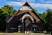
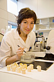
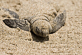
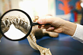
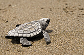
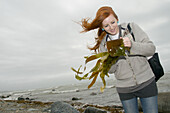
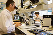
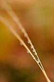
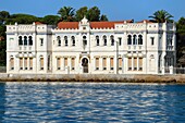
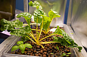
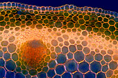
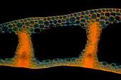
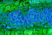
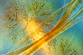
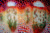
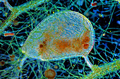
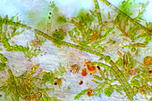
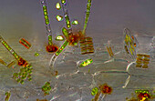
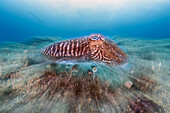
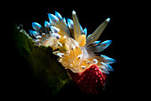
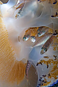
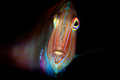
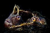
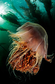
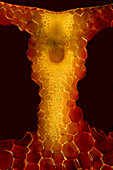
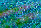
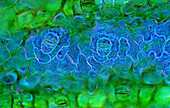
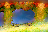
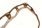
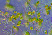
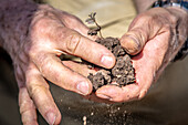
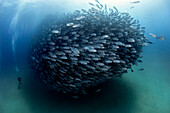
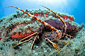
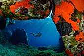
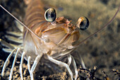
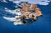
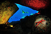
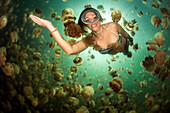
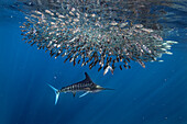
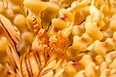
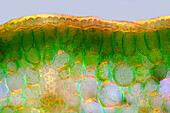
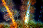
71187984 - Biology Museum on Djurgarden, Stockholm, Sweden70208462 - Microtome, Fishing biology laboratory. Polishing and reading of otoliths and gonads. AZTI-Tecnalia. Technological Centre specialised in Marine and Food Research. Pasaia, Gipuzkoa, Euskadi. Spain.70286310 - Baby, Biology, Born, Close-up, Color, Colour, Conservation, Ecology, Horizontal, Life, Lora, Marine, Marine life, Mexico, Rising, Sand, Shore, Tecolutla, Turtle, Veracruz, Wild, V03-839683, agefotostock 70374771 - Canada, Québec, Montreal, private school, biology class70286311 - Baby, Biology, Born, Close-up, Color, Colour, Conservation, Ecology, Horizontal, Life, Lora, Marine, Marine life, Mexico, Rising, Sand, Shore, Tecolutla, Turtle, Veracruz, Wild, V03-839688, agefotostock 70286312 - Baby, Biology, Born, Close-up, Color, Colour, Conservation, Ecology, Horizontal, Life, Lora, Marine, Marine life, Mexico, Rising, Salt, Sand, Sea, Shore, Swim, Tecolutla, Turtle, Veracruz, Water, Wild, V03-839692, agefotostock 70255851 - young teenagewoman on marine biology field course studying kelp seaweed on the beach, Criccieth, North Wales, windy afternoon70208460 - Fishing biology laboratory. Polishing and reading of otoliths and gonads. AZTI-Tecnalia. Technological Centre specialised in Marine and Food Research. Pasaia, Gipuzkoa, Euskadi. Spain.70395770 - Close up of dropper in petri dish. Biological research. Liquid being pipetted into a petri dish during experiment used in DNA, microbiology, biology, genetics,biomedical and pharma research70384434 - America, backdrop, background, beautiful, beauty, bend, bending, biology, botanical, botany, brown, Canada, Canadian, clean, Close-up, Color image, colourful, Copy space, country, daydreaming, delicate, detail, dream, dreamer, ecology, ecosystem, elegant,70384433 - America, backdrop, background, beautiful, beauty, bend, bending, biology, botanical, botany, brown, Canada, Canadian, clean, Close-up, Color image, colourful, Copy space, country, daydreaming, delicate, detail, dream, dreamer, ecology, ecosystem, elegant,70208461 - Fishing biology laboratory. Polishing and reading of otoliths and gonads. AZTI-Tecnalia. Technological Centre specialised in Marine and Food Research. Pasaia, Gipuzkoa, Euskadi. Spain.14097820 - France, Var, the Rade (Roadstead) of Toulon, La Seyne sur Mer, area of Tamaris, the orientalist style Michel Pacha Institute (formerly Institute of Marine Biology of the University of Lyon) and the Crescent villa (villa du Croissant) is recognizable by its minaret tower14100194 - France, Var, the Rade (Roadstead) of Toulon, La Seyne sur Mer, area of Tamaris, the orientalist style Michel Pacha Institute (formerly Institute of Marine Biology of the University of Lyon)14127312 - Aquaponics at College Park UMD13925580 - The image presents Anemone sylvestris stalk in transversal cross-section, photographed through the microscope in polarized light at a magnification of 200X\n13925525 - The image presents Carex sp. leaf in transversal cross-section, photographed through the microscope in polarized light at a magnification of 100X\n13925473 - The image presents stomata in Spathiphyllum leaf epidermis, photographed through the microscope in polarized light at a magnification of 100X\n13925385 - The image presents Fragilaria sp., a kind of diatoms against Batrachospermum, a kind of red algae, photographed through the microscope in polarized light at a magnification of 200X\n13925332 - The image presents stomata in hosta leaf epidermis, photographed through the microscope in polarized light at a magnification of 100X\n13925278 - The image presents various tiny algae settled on Lemna sp. root, photographed through the microscope in polarized light at a magnification of 400X. On the right are visible diatoms closed in a special protecting case.\n13925207 - The image presents stomata in Spathiphyllum leaf epidermis, photographed through the microscope in polarized light at a magnification of 400X\n13925148 - The image presents stomata in Spathiphyllum leaf epidermis, photographed through the microscope in polarized light at a magnification of 400X\n13925085 - The image presents vascular bundles in senecio stalk, photographed through the microscope in polarized light at a magnification of 200X\n13925083 - The image presents Utricularia trap, a kind of carnivorous plant, photographed through the microscope in polarized light and dark field, at a magnification of 100X\n13925044 - The image presents various tiny algae settled on Lemna sp. root, photographed through the microscope in polarized light at a magnification of 200X\n13925041 - The image presents various tiny algae settled on Lemna sp. root, photographed through the microscope in polarized light at a magnification of 200X\n13925010 - The image presents read leaf in transversal cross-section, photographed through the microscope in polarized light at a magnification of 100X\n13924337 - The image presents tissues in nettle stalk in longitudinal cross section, photographed through the microscope in polarized light at a magnification of 100X\n13847358 - A hand holds a rock up for examination in a field near Debre Berhan, Ethiopia13846345 - Local Ethiopian farmers look on as a field researcher examines the soil of a hillside, Debre Berhan, Ethiopia.13845483 - Hands break up chunks of soil, Debre Berhan, Ethiopia.13845468 - The Shark Reef Aquarium at Mandalay Bay hotel and casino in Las Vegas. The Shark Reef Aquarium is comprised of nearly 1.6 million gallons of water.13845061 - Close up look at female scientist analyzing petri dish containing samples of peanut plant roots, Tifton, Georgia.13821242 - Dreamlike shot of a humpback whale (Megaptera novaeangliae) in the waters of Tonga island (south Pacific)13821237 - A Mediterranean slipper lobster (Scyllarides latus) during a night dive13821229 - A common cuttlefish in a zoomed portrait13821227 - Clos up of one of the most beautiful nudibranchs in the Mediterranean, the Antiopella cristata, Italy13821225 - Juvenile Atlantic horse mackerel fishes (Trachurus trachurus) find refuge in the lobes of a mediterranean fried egg jellyfish (Cotylorhiza tuberculata), a typical symbiosis of our seas.13821224 - Close of portrait of a Pearly Razorfish or Cleaver Wrasse (Xyrichtys novacula), one of the most shy and elusive fish in the Mediterranean, Italy13821222 - A couple of butterfly blenny (Blennius ocellaris) peeps out from inside a fan mussel (Pinna nobilis), that they chose as the nest for hatching their eggs.13821215 - Sea bottom of False Bay are the natural habitat of sharks, seals, whales and dolphins, South Africa13821210 - A beautiful Godiva quadricolor nudibranch lays its eggs on a plastic bottle.13821209 - A nursehound (Scyliorhinus stellaris) watches closely a group of small cod passing by. Shot taken off Chioggia (VE) on one of the many rocky formations developed in the seabed of the upper Adriatic, called Tegnue. These bio-sediments are similar to coral reefs and for this reason they are also called Adriatic coral reefs.13821208 - A small cuttlefish (Sepia officinalis) still in its egg, about 1 cm whole size. Eyes, head and tentacles are already perfectly formed and defined, this means that only a few days are left to hatch, although the little cephalopod is still connected to its yolk sac. This picture is part of a work that features a special Italian place, Numana, on the Adriatic sea, where very specific geographical conditions make this spot a realm for underwater macro subjects, pretty much like Philippines or Indonesia.13821196 - An iconic shot of a very common mediterranean subject, the red sea fan (Paramuricea clavata), Italy13821193 - A beautiful mediterranean nudibranch (Spurilla neapolitana). This picture is part of a work started a few years ago to feature a special italian place, Numana, on the Adriatic sea, where very specific geographical conditions make this spot a realm for underwater macro subjects, pretty much like Philippines or Indonesia.13821185 - A beautiful glimpse of a deep Mediterranean landscape with black coral (Antipathella subpinnata), red sea-fan (Paramuricea clavata) and basket star (Astrospartus mediterraneus). Formiche di Grosseto (Tuscan Archipelago)13821183 - Lion's mane jellyfish (Cyanea capillata) in a kelp forest at God's Pocket Marine Park, Port Hardy, British Columbia, Canada13925579 - The image presents stomata in Spathiphyllum leaf epidermis, photographed through the microscope in polarized light at a magnification of 200X\n13925423 - The image presentstwo suctorians ( a kind of ciliate) and tiny diatoms, photographed through the microscope in polarized light at a magnification of 200X\n13925392 - The image presents reed stalk in transversal cross-section, photographed through the microscope in polarized light at a magnification of 200X\n13925367 - The image presents Ophrydium sp. ( a kind of colonial ciliates), photographed through the microscope in polarized light at a magnification of 100X\n13925287 - The image presents tissues in nettle stalk in longitudinal cross-section, photographed through the microscope in polarized light at a magnification of 100X\n13925265 - The image presents nettle tissues in the stalk in longitudinal cross-section, photographed through the microscope in polarized light at a magnification of 100X\n13925104 - The image presents diptera eggs, photographed through the microscope in polarized light at a magnification of 100X\n13925100 - The image presents stomata in Stromanthe sp. leaf epidermis, photographed through the microscope in polarized light at a magnification of 100X\n13925093 - The image presents a single vascular bundle in Carex sp. stalk, photographed through the microscope in polarized light and dark field at a magnification of 200X\n13925078 - The image presents stomata in lily leaf epidermis, photographed through the microscope in polarized light at a magnification of 200X\n13925070 - The image presents stomata in Spathiphyllum sp. leaf epidermis, photographed through the microscope in polarized light at a magnification of 100X\n13925038 - The image presents stomata in Spathiphyllum leaf epidermis, photographed through the microscope in polarized light at a magnification of 200X\n13924900 - The image presents stomata in Spathiphyllum sp. leaf epidermis, photographed through the microscope in polarized light at a magnification of 100X\n13924894 - The image presentstwo suctorians ( a kind of ciliate) and tiny diatoms, photographed through the microscope in polarized light at a magnification of 200X\n13924866 - The image presents reed stalk in transversal cross-section, photographed through the microscope in polarized light at a magnification of 200X\n13924859 - "The image presents Cladophora sp. ""twigs"" (a kind of green algae) with Cocconeis sp. (a kin of diatoms) settled on it, photographed through the microscope in polarized light at a magnification of 200X"\n13924803 - The image presents Fragilaria sp., a kind of diatoms against Batrachospermum, a kind of red algae, photographed through the microscope in polarized light at a magnification of 200X\n13924786 - The image presents knautia arvensis tissues in the transversal section of the stalk, photographed through the microscope in bright field, at a magnification of 100X\n13924785 - The image presents diatoms (mostly Gomphonema sp.) photographed through the microscope in polarized light at a magnification of 200X\n13924764 - The image presents Batrachospermum sp., a kind of red algae, photographed through the microscope in polarized light at a magnification of 200X\n13924748 - The image presents a single stoma in Knautia arvensis epidermis, photographed through the microscope in polarized light at a magnification of 200X\n13924602 - The image presents Fragilaria sp., a kind of diatoms and Batrachospermum sp., a kind of red algae, photographed through the microscope in polarized light at a magnification of 200X\n13924555 - The image presents tissues in nettle stalk in longitudinal cross-section, photographed through the microscope in polarized light at a magnification of 100X\n13924552 - The image presents stomata in Spathiphyllum leaf epidermis, photographed through the microscope in polarized light at a magnification of 400X\n13924513 - The image presents various tiny algae settled on Lemna sp. root, photographed through the microscope in polarized light at a magnification of 200X\n13924452 - The image presents reed stalk in transversal cross-section, photographed through the microscope in polarized light at a magnification of 200X\n13924434 - The image presents carex sp. leaf in transversal cross-section, photographed through the microscope in polarized light at a magnification of 100X\n13924404 - The image presents euglenoids among green algae, photographed through the microscope in polarized light at a magnification of 100X\n13848999 - Hands break up chunks of soil, Debre Berhan, Ethiopia.13848106 - Close up look hand holding petri dish containing samples of peanut plant roots, Tifton, Georgia.13845943 - Young Asian-American woman concentrates on food science experiment, College Park, Maryland13845918 - Researcher in a lab studying a sample13844386 - Morel mushrooms of Fairbanks Alaska13824447 - Hiker shows wildlife notes about Pato Cortacorrientes, Merganetta armata, near Chileno refuge, Torres del Paine national park, Patagonia, Chile13821356 - A diver admires in awe a big aggregation of jack fish in the waters of Cabo Pulmo Marine National Park, where marine biomass has increased exponentially since the marine park was established in 199513821230 - A spiny lobster (Palinurus elephas) rests inside a small cave close to surface, Italy13821220 - Diver in a nice cave of Capraia island, Tuscan archipelago.13821219 - Close up portrait of a shrimp (Penaeus kerathurus) in Italy13821218 - A exceptional and very rare encounter with the sea lamprey (Petromyzon marinus) in Italy13821217 - A beautiful Caneva's blenny (Lipophrys canevae) peeps out of its burrow13821213 - A sand tiger shark (Carcharias taurus) in the famous cave of Aliwal Shoal, where in the months of the South African winter these sharks meet in large numbers for mating and reproduction.13821212 - The mythical cow shark or “seven gills” (Notorhyncus cepedianus), the only existing example of the genus Notorhyncus of the Hexanchidae family, is easily recognized by the presence of 7 gill slits, typical of sharks of the ancient Mesozoic species.13821207 - A sea turtle (Caretta caretta) striving to get free from a plastic fishing net in Spain13821206 - Diver and artistic lighting shot of the cave of Scilla, Italy13821204 - A smiling female diver among the golden jellyfish (Mastigias papua) of Jellyfish Lake, on the island of Eil Malk (Republic of Palau, Micronesia).13821203 - A striped marlin (Kajikia audax) chases a group of very fast mackerel (Scomber Disbrus) in the waters of Magdalena Bay, off the village of Puerto San Carlo, Baja California Sur, Mexico13821201 - A diver in the dense marsh vegetation of the Corrego Azul river, close to Bonito, Mato Grosso do Sul, Brazil13776355 - A close look at a squat shrimp (Thor amboinensis), on a beaded anemone (Heteractis aurora). This anemone has been found as host to 7 different species of clownfish as well as the domino damselfish; Philippines13925468 - The image presents palisade mesophyll in hyacinthus leaf (transversal cross-section) photographed through the microscope in polarized light at a magnification of 200X\n13925399 - The image presents Batrachospermum sp., a kind of red algae, and FRagilaria sp., a kind of diatoms, photographed through the microscope in polarized light and dark field at a magnification of 100X\nnext page